Buriak and her team have designed nanoparticles that absorb light and conduct electricity from two very common elements: phosphorus and zinc. Both materials are more plentiful than scarce materials such as cadmium and free from manufacturing restrictions imposed on lead-based nanoparticles."Half the world already lives off the grid, and with demand for electrical power expected to double by the year 2050, it is important that renewable energy sources like solar power are made more affordable by lowering the costs of manufacturing," Buriak said.Her team's research supports a promising approach of making solar cells cheaply using mass manufacturing methods like roll-to-roll printing (as with newspaper presses) or spray-coating (similar to automotive painting). "Nanoparticle-based 'inks' could be used to literally paint or print solar cells or precise compositions," Buriak said.The team was able to develop a synthetic method to make zinc phosphide nanoparticles and demonstrated that the particles can be dissolved to form an ink and processed to make thin films that are responsive to light.Buriak and her team are now experimenting with the nanoparticles, spray-coating them onto large solar cells to test their efficiency. The team has applied for a provisional patent and has secured funding to enable the next step to scale-up manufacture.
Saturday, August 31, 2013

Friday, September 16, 2011
Diet food making you fat

THE idea of an ice cream that is lighter, sugar free or has low/ no fat was every dieters dream, which ice cream manufacturers didn’t take long to translate into reality. Amul's Slim Scoop, Diet Scoop, Dairy Den, Havmor's Vanilla, or Vadilal’s or Mother Diary's Dietz are some of the ice creams marketed as diet products.
DIET CHOCOLATES
FAT and sugar free chocolates are a dream come true for chocoholics on a weight loss regime. There are also fat free chocolate pudding cups, which claim to have only 35 calories compared to 80 calories in the regular pudding. However, experts have a different take on this.
"Don't get into the trap of no-fat chocolates. When manufacturers remove the sweet stuff, they add fat. These might also contain the element, sorbitol, which can cause health problems like bloating and diarrhea.
LOW FAT BUTTER
CONSUMPTION of low fat butter is of course any day a better choice than the regular stuff but too much of that can make you fat too. Nutralite and Amul Lite, the low calorie bread spreads are a popular choice among the health conscious. These are cholesterol free, low in saturated fats.
DIET SNACKS
GO TO any of the urban supermarkets, and you won’t miss those ubiquitous fat free sweets, jams, muffins and bhujias . All these, promising you to take on guilt free trip! But beware, as most of these snacks are also sugar and fat bombs in disguise.
Gorge on them, yes, but not with false hopes, warn experts.
Food Types for Healthy Hair
When your hair starts falling off like dry straws, what can you do?
First, it would help to locate the various reasons for hair loss. Studies have revealed that men lose hair for some common reasons, like:
- Your scalp needs nutrition. So get plenty of fluids like buttermilk, lime juice and coconut water. Drink at least 8-10 glasses of water every day!
- One bowl of sprouted pulses for would be great.
- Proteins. Eat chicken and egg each day.
- Reduce you daily intake of tea and coffee.
- When you are washing hair, use a teaspoon of lime juice for the last rinse. That make your hair lustrous while protecting it from dandruff.
- You are never too old for milk. Drink two glasses of skimmed milk for maximum benefits.
- Apply a paste of methi seeds on your scalp for half an hour once every week, and drink methi water on empty stomachs.
- Remember how the age old sadhus survived on fruits? No wonder that they had such long hair! Eat at least 2 or 3 fruits every day like strawberries, bananas, apples, mangoes, chikoos, and grapes.
- Go green- eat a lot of vegetables, munch away on that iron laden spinach.
- It is better to have more faith in white meat. Fish, chicken and egg can do wonders for your hair! Try to stay away from red meat.
Five Healthy Foods to Eat at your Workstation
Not only they help you conquer that “false hunger”, they are also brimming with nutrients which are needed to keep you going all day. You can choose between apples, bananas, mangoes, oranges, papayas etc. but make sure you carry some fruits to the office on a daily basis.

Cup of Green Tea
Polyphenols, one of the major ingredients of green tea, is known to help repair the damaged DNA and stimulate the immune system. Besides, research shows that green tea also helps in reducing the risk of cancers, including skin cancer and prostate cancer.

Walnuts
Some new researches have pointed out that walnuts have more (and better quality) antioxidants than almonds, peanuts, pistachios and many other nuts. All nuts have been found to improve lipids and can reduce the risk of heart diseases. Walnuts have also been found to enhance cognitive and motor function in animals with Alzheimer's. Experts recommend seven walnuts a day.

Popcorn
If there ever was a list of healthy snacks, this is definitely going to be on the list. In fact, many say that popcorn is the healthiest snack available. It is 100 percent whole grain and is big on fiber and antioxidants. If you can air-pop your popcorn and add a minimal quantity of salt, you'd have the best popcorn, say experts. If your office has a microwave, just make sure you bring a pack of low sodium popcorn to work.

Dark Chocolate
This, too, contains those powerful antioxidants called Polyphenols which combat the risk factors (like oxidation of LDL cholesterol) for heart diseases and also inhibit clotting. Studies have also indicated that consuming a small bar of dark chocolate daily can reduce blood pressure in people with mild hypertension. Caffeine, another active ingredient of dark chocolate, can give you that much-required kick and a boost in concentration levels.

Thursday, September 8, 2011
Stem Cell Assays
Stem cell therapy development is an exciting area of research that promises the future treatment of many diseases. Since this area in drug development is in its infancy, there are many hurdles to overcome. The scientists at Marin Biologic are here to assist you in the many phases of your stem cell therapy development. We can culture adult or embryonic stem cells and monitor the differentiation markers for your therapy development. Please think of Marin Biologic as your stem cell research laboratory.
Microenvironment may contribute to stem cell "homing" events or differentiation. Our scientists can study microenvironments, whether generated in culture or from exogeneous tissue, and their effects on stem cell differentiation using in vitro methods. Transfecting adult or embryonic stem cells with a genetic marker to follow their "homing activities" can be performed prior to inoculation in vivo. Development of "homing" stem cells as a delivery vehicle for cancer therapy can be undertaken at Marin Biologic. Replacement therapy for hormone, cytokines and metabolic intermediates can be implemented using stem cell therapy. Insulin production is a key target for diabetes therapy using stem cells because of their "normal" detection of glucose levels and "normal" synthesis and release of insulin. Our scientists can help you to develop stems cells that synthesize your therapeutic molecule by initiating differentiation and monitoring specific production.
To implement stem cell therapy, there must be many levels of investigation ranging from the cell culture of stem cells, triggering differentiation, expanding stem cell cultures to large scale cultures for replacement therapy, determining whether immunocompatibility of embryonic stem cells is retained for transplant, down regulating HLA molecules in adult stem cells, etc. The scientists at Marin Biologic are very experienced and creative in cell biology and can bridge this field with molecular biology and biochemistry for potential cell therapy and assay development.
Microenvironment may contribute to stem cell "homing" events or differentiation. Our scientists can study microenvironments, whether generated in culture or from exogeneous tissue, and their effects on stem cell differentiation using in vitro methods. Transfecting adult or embryonic stem cells with a genetic marker to follow their "homing activities" can be performed prior to inoculation in vivo. Development of "homing" stem cells as a delivery vehicle for cancer therapy can be undertaken at Marin Biologic. Replacement therapy for hormone, cytokines and metabolic intermediates can be implemented using stem cell therapy. Insulin production is a key target for diabetes therapy using stem cells because of their "normal" detection of glucose levels and "normal" synthesis and release of insulin. Our scientists can help you to develop stems cells that synthesize your therapeutic molecule by initiating differentiation and monitoring specific production.
To implement stem cell therapy, there must be many levels of investigation ranging from the cell culture of stem cells, triggering differentiation, expanding stem cell cultures to large scale cultures for replacement therapy, determining whether immunocompatibility of embryonic stem cells is retained for transplant, down regulating HLA molecules in adult stem cells, etc. The scientists at Marin Biologic are very experienced and creative in cell biology and can bridge this field with molecular biology and biochemistry for potential cell therapy and assay development.
CELL BASED ASSAY
Cell-based assay or bioassay development can range from cytotoxic assays including apoptosis to cell proliferation and metabolic assays. Cell-based assay development can also include high throughput screening assays and other custom bioassays used to characterize drug stability for GLP GMP lot release, drug potency and for drug purification and production support. Mechanisms of action, such as receptor binding, receptor activation, cell signaling, drug internalization and subcellular localization can be delineated in cell-based assays following treatment with your pharmaceuticals. Bioassay development can encompass testing of conditioned medium, cell lysates or whole cells in a variety of formats including ELISA and immunohistochemical methods. Our scientists can grow and differentiate stem cells as well as provide assay development to monitor stem cell differentiation using specific gene expression.
Sunday, December 26, 2010
Human Genome (DNA) by Numbers (statistics)
Cells in the human bodY - 75-100 trillion Base pairs in each cell -3.1 billion
Base pairs in the largest human gene (dystrophin) -2.4 million
Genes in the human genome - 28,000-35,000
Chromosomes in each cell - 46
Human DNA measurements: DNA helix diameter - 20-26 Ångström2.0-2.6 Nanometers
Distance between base pairs - 3.3-3.4 Ångström0.33-0.34 Nanometers
Length of one helix turn - 33-34 Ångströమ - 3.3-3.4 Nanometers
Number of base pairs in one helix turn - 10
How Human DNA compares to other species:
Below we listed a number of species and their DNA properties like number of chromosomes, genes and base pairs and how they differentiate in comparison to human DNA.
species CHROMOSOMES GENES BASEPAIRS
Human (Homo sapiens)
46 (23 pairs)
23,686
~3.1 billion
Mouse (Mus musculus)
40
23,786
~2.7 billion
Pufferfish (Takifugu rubripes)
44
~31,000
~365 million
Malaria Mosquito (Anopheles gambiae)
6
~14,000
~289 million
Sea Squirt (Ciona intestinalis)
28
~16,000
~160 million
Fruit Fly (Drosophila melanogaster)
8
~14,000
~137 million
Roundworm (Caenorhabditis elegans)
12
~19,000
~97 million
Bacterium (Escherichia coli)
(1)
~5,000
~4.1 million
ANIMAL
Percentage of human DNA
Chimpanzee
96.0%
Orang Utan
96.4%
Gorilla
97.7%
Bonobo
98.4%
Mice
70-98.5%
The Human DNA Structure
The Human DNA (Deoxyribonucleic acid) consists of more than 3 billion base pairs of 4 different nucleic acids (nucleotides) that make up the genetic code of a human. This equals about 750 Megabytes of data that holds all instructions needed for the development of a complete living organism. It is therefore often referred to as a "blueprint" to construct all cells, organs, skin, hair, nails and other components (such as proteins and RNA) necessary for creating a functioning organism like our body. This genetic information is stored in segments of the DNA called "genes". This is the main purpose of DNA - the long-term storage of genetic information. The DNA contained in Human Embryonic Stem Cells can let them grow into every part of the human body such as heart, liver, kidney, brain, ears, eyes, bones or skin. As such, these cells are currently subject of intensive scientific research. The results could provide many new possibilities for therapeutically treatments for a whole range of diseases that are currently deemed incurable.

The DNA resides in the nucleus of each cell in the human body. Inside the nucleus of each body cell, there are 46 Chromosomes in humans, which is 2 complete identically sets of 22 chromosomes (autosome chromosomes) and 2 additional gender specific chromosomes (sex chromosomes) - either 2 X chromosomes (XX) for a female (woman) or 1 X and 1 Y chromosome (XY) for a male (man). Therefore humans are considered diploid as each body cell contains 2 homologous copies of each chromosome, normally one from the mother and one from the father. However, human haploid gametes (sperm and egg) only contain 23 chromosomes - haploid cells and organisms only have 1 copy of chromosomes. These 46 chromosomes are nothing else than an organized structure of the coiled human DNA and certain proteins.
The shape of DNA describes a long spiral, comparable to a twisted rope ladder or a spiral staircase with a fixed diameter. Also called a "double helix", the spiral forming the human DNA molecule is chemically made up of 2 strands of a sugar-phosphate backbone, running antiparallel. This backbone (skeleton) consists of a phosphate group (Phosphoric Acid, H3PO4) and a sugar group (Deoxyribose, C5H10O4) forming phosphodiester bonds between them, resulting in the Phosphate-Deoxyribose backbone. In between those 2 twisted strands of DNA there are complementary purine-pyrimidine base pairs holding the two strands. There are only 2 possible base pairs combinations:
Adenine (C5H5N5) and Thymine (C5H6N2O2)
Guanine (C5H5N5O) and Cytosine (C4H5N3O)
Those base pairs forming 2 (Adenine-Thymine) or 3 (Guanine-Cytosine) hydrogen bonds between each other. They are connected to the sugar molecule of the 2 backbone strands. The complex of a sugar-phosphate molecule together with a base molecule forms a "nucleotide". The chemical formula of Adenine, Thymine, Guanine and Cytosine (their molecular formula) is very similar and explains the binding process. The DNA structure model or double helix model of human DNA as we know it today (twisted ladder model or spiral staircase) was discovered in 1953 by James D. Watson and Francis Crick.
Human DNA Sequence
The human DNA sequence is unique for every individual person, even though it is nearly 99.9% identical. It's the tiny portion of only 0.1% of DNA that differentiates us from every other human and that contributes to our individual differences. These small variations in the human genome such as "Single Nucleotide Polymorphisms" (SNPs) and the "Variable Number Tandem Repeat" (VNTR) allow further analysis using DNA fingerprinting (DNA profiling) techniques. The results of this kind of human DNA analysis are being used for ancestry testing, paternity testing or especially for forensic criminal investigations. The later shows the increasingly important role of genetics in investigating crimes such as rape or murder; hence human DNA can act as evidence in court cases.

The DNA resides in the nucleus of each cell in the human body. Inside the nucleus of each body cell, there are 46 Chromosomes in humans, which is 2 complete identically sets of 22 chromosomes (autosome chromosomes) and 2 additional gender specific chromosomes (sex chromosomes) - either 2 X chromosomes (XX) for a female (woman) or 1 X and 1 Y chromosome (XY) for a male (man). Therefore humans are considered diploid as each body cell contains 2 homologous copies of each chromosome, normally one from the mother and one from the father. However, human haploid gametes (sperm and egg) only contain 23 chromosomes - haploid cells and organisms only have 1 copy of chromosomes. These 46 chromosomes are nothing else than an organized structure of the coiled human DNA and certain proteins.
The shape of DNA describes a long spiral, comparable to a twisted rope ladder or a spiral staircase with a fixed diameter. Also called a "double helix", the spiral forming the human DNA molecule is chemically made up of 2 strands of a sugar-phosphate backbone, running antiparallel. This backbone (skeleton) consists of a phosphate group (Phosphoric Acid, H3PO4) and a sugar group (Deoxyribose, C5H10O4) forming phosphodiester bonds between them, resulting in the Phosphate-Deoxyribose backbone. In between those 2 twisted strands of DNA there are complementary purine-pyrimidine base pairs holding the two strands. There are only 2 possible base pairs combinations:
Adenine (C5H5N5) and Thymine (C5H6N2O2)
Guanine (C5H5N5O) and Cytosine (C4H5N3O)
Those base pairs forming 2 (Adenine-Thymine) or 3 (Guanine-Cytosine) hydrogen bonds between each other. They are connected to the sugar molecule of the 2 backbone strands. The complex of a sugar-phosphate molecule together with a base molecule forms a "nucleotide". The chemical formula of Adenine, Thymine, Guanine and Cytosine (their molecular formula) is very similar and explains the binding process. The DNA structure model or double helix model of human DNA as we know it today (twisted ladder model or spiral staircase) was discovered in 1953 by James D. Watson and Francis Crick.
Human DNA Sequence
The human DNA sequence is unique for every individual person, even though it is nearly 99.9% identical. It's the tiny portion of only 0.1% of DNA that differentiates us from every other human and that contributes to our individual differences. These small variations in the human genome such as "Single Nucleotide Polymorphisms" (SNPs) and the "Variable Number Tandem Repeat" (VNTR) allow further analysis using DNA fingerprinting (DNA profiling) techniques. The results of this kind of human DNA analysis are being used for ancestry testing, paternity testing or especially for forensic criminal investigations. The later shows the increasingly important role of genetics in investigating crimes such as rape or murder; hence human DNA can act as evidence in court cases.
How Larger DNA Fragments Can Be Cloned?
Both λ phage vectors and the more commonly used E. coli plasmid vectors are useful for cloning DNA fragments up to ≈20 – 25 kb. However, cloning of much larger fragments is desirable for sequencing of extremely long DNAs such as the DNA in a eukaryotic chromosome. Also, because of the common occurrence of large introns in genes from higher eukaryotes, it is often necessary to clone DNA fragments greater than 25 kb in order to include an entire gene in one clone. Consequently, additional types of cloning vectors have been developed for cloning larger fragments of DNA.
One common method for cloning larger fragments makes use of elements of both plasmid and λ phage cloning. In this method, called cosmid cloning, recombinant plasmids containing inserted fragments up to 45 kb long can be efficiently introduced into E. coli cells. A cosmid vector is produced by inserting the COS sequence from λ phage DNA into a small E. coli plasmid vector about 5 kb long. Like other plasmid vectors discussed earlier, cosmid vectors contain a replication origin (ORI), an antibiotic-resistance gene (e.g., ampr), and a polylinker sequence containing numerous restriction-enzyme recognition sites.Next, the cosmid vector is cut with a restriction enzyme and then ligated to 35- to 45-kb restriction fragments of foreign DNA with complementary sticky ends. If the concentration of foreign DNA is high enough, the ligation reaction generates long DNA molecules containing multiple restriction fragments of the foreign DNA separated by the 5-kb cosmid DNA. These ligated DNA molecules, which resemble the concatomers that form during replication of λ phage in a host cell, can be packaged in vitro as described earlier.

General procedure for cloning DNA fragments in cosmid vectors. This procedure has the high efficiency associated with λ phage cloning and permits cloning of restriction fragments.
In the packaging reaction, the λ Nu1 and A proteins bind to COS sites in the ligated DNA and direct insertion of the DNA between two adjacent COS sites into empty phage heads. Packaging will occur so long as the distance between adjacent COS sites does not exceed about 50 kb (the approximate size of the λ genome). Phage tails then are attached to the filled heads, producing viral particles that contain a recombinant cosmid DNA molecule rather than the λ genome. When these virions are plated on a lawn of E. coli cells, they bind to phage receptors on the cell surface and inject the packaged DNA into the cells.
Since the injected DNA does not encode any λ proteins, no viral particles form in infected cells and no plaques develop on the plate. Rather, the injected DNA forms a large circular plasmid, composed of the cosmid vector and an inserted DNA fragment, in each host cell. This plasmid replicates and is segregated to daughter cells like other E. coli plasmids and the colonies that arise from transformed cells can be selected on antibiotic plates. The high efficiency of λ phage infection of E. coli cells makes cosmid cloning a practical method of generating plasmid clones carrying DNA fragments up to 45 kb long. Since many genes of higher eukaryotes are on the order of 30 – 40 kb in length, cosmid cloning increases the chances of obtaining DNA clones containing the entire sequences of genes.
One common method for cloning larger fragments makes use of elements of both plasmid and λ phage cloning. In this method, called cosmid cloning, recombinant plasmids containing inserted fragments up to 45 kb long can be efficiently introduced into E. coli cells. A cosmid vector is produced by inserting the COS sequence from λ phage DNA into a small E. coli plasmid vector about 5 kb long. Like other plasmid vectors discussed earlier, cosmid vectors contain a replication origin (ORI), an antibiotic-resistance gene (e.g., ampr), and a polylinker sequence containing numerous restriction-enzyme recognition sites.Next, the cosmid vector is cut with a restriction enzyme and then ligated to 35- to 45-kb restriction fragments of foreign DNA with complementary sticky ends. If the concentration of foreign DNA is high enough, the ligation reaction generates long DNA molecules containing multiple restriction fragments of the foreign DNA separated by the 5-kb cosmid DNA. These ligated DNA molecules, which resemble the concatomers that form during replication of λ phage in a host cell, can be packaged in vitro as described earlier.

General procedure for cloning DNA fragments in cosmid vectors. This procedure has the high efficiency associated with λ phage cloning and permits cloning of restriction fragments.
In the packaging reaction, the λ Nu1 and A proteins bind to COS sites in the ligated DNA and direct insertion of the DNA between two adjacent COS sites into empty phage heads. Packaging will occur so long as the distance between adjacent COS sites does not exceed about 50 kb (the approximate size of the λ genome). Phage tails then are attached to the filled heads, producing viral particles that contain a recombinant cosmid DNA molecule rather than the λ genome. When these virions are plated on a lawn of E. coli cells, they bind to phage receptors on the cell surface and inject the packaged DNA into the cells.
Since the injected DNA does not encode any λ proteins, no viral particles form in infected cells and no plaques develop on the plate. Rather, the injected DNA forms a large circular plasmid, composed of the cosmid vector and an inserted DNA fragment, in each host cell. This plasmid replicates and is segregated to daughter cells like other E. coli plasmids and the colonies that arise from transformed cells can be selected on antibiotic plates. The high efficiency of λ phage infection of E. coli cells makes cosmid cloning a practical method of generating plasmid clones carrying DNA fragments up to 45 kb long. Since many genes of higher eukaryotes are on the order of 30 – 40 kb in length, cosmid cloning increases the chances of obtaining DNA clones containing the entire sequences of genes.
cDNA Libraries Are Prepared from Isolated mRNAs
In higher eukaryotes, many genes are transcribed into mRNA only in specialized cell types. For example, mRNAs encoding globin proteins are found only in erythrocyte precursor cells, called reticulocytes. Likewise, the mRNA encoding albumin, the major protein in serum, is produced only in liver cells where albumin is synthesized. The specific DNA sequences expressed as mRNAs in a particular cell type can be cloned by synthesizing DNA copies of the mRNAs isolated from that type of cell, and then cloning the DNA copies in plasmid or bacteriophage λ vectors.
DNA copies of mRNAs are called complementary DNAs(cDNAs); clones of such DNA copies of mRNAs are called cDNA clones. In addition to representing only the sequences expressed as mRNAs in a particular cell type, cDNA clones lack the noncoding introns present in genomic DNA clones. Thus the amino acid sequence of a protein can be determined directly from the nucleotide sequence of its corresponding cDNA. Many genes in higher eukaryotes are too large to be included in a single λ clone because of their large introns. In contrast, all full-length cDNAs, containing the entire protein-coding sequence, can be included in a single λ clone. However, because of methodological difficulties, not all cDNA clones are full length when initially produced; to obtain a full-length cDNA, it often is necessary to isolate several overlapping cDNA clones and then ligate them at rare restriction sites. Just as a large collection of clones containing fragments of genomic DNA representing the entire genome of a species is called a genomic library, a large collection of cDNA copies of all the mRNAs in a cell type is called a cDNA library.
Isolation of mRNAs and Synthesis of cDNAs
The first step in preparing a cDNA library is to isolate the total mRNA from the cell type or tissue of interest. Nature has greatly simplified the isolation of eukaryotic mRNAs: the 3′ end of nearly all eukaryotic mRNAs consists of a string of 50 – 250 adenylate residues, the poly(A) tail. Because of their poly(A) tail, mRNAs can be easily separated from the much more prevalent rRNAs and tRNAs present in a cell extract by use of a column to which short strings of thymidylate (oligo-dTs) are linked to the matrix. When a cell extract is passed through an oligo-dT column, the mRNA poly(A) tails base-pair with the oligo-dTs, binding the mRNAs to the column. Since rRNAs, tRNAs, and other molecules do not bind to the column, they can be washed away. The bound mRNAs are recovered by elution with a low-salt buffer.

Isolation of eukaryotic mRNA by oligo-dT column affinity chromatography. Isolated cytoplasmic RNA consists mostly of ribosomal RNAs (rRNAs) and transfer RNAs (tRNAs).
The enzyme reverse transcriptase, which is found in retroviruses, is then used to synthesize a strand of DNA complementary to each mRNA molecule. This enzyme can polymerize deoxynucleoside triphosphates into a complementary DNA strand using an RNA molecule as template. Like other DNA polymerases, reverse transcriptase can add nucleo-tides only to the 3′ end of a preexisting primer base-paired to the template. Added free oligo-dT serves this function by hybridizing to the 3′ poly(A) tail of each mRNA template.

Preparation of a bacteriophage λ cDNA library. A mixture of mRNAs, isolated as shown in Figure is used to produce cDNAs corresponding to all the cellular mRNAs.
Conversion of Single-Stranded cDNA to Double-Stranded cDNA
After cDNA copies of isolated mRNAs are synthesized, the mRNAs are removed by treatment with alkali, which hydrolyzes RNA but not DNA. The single-stranded cDNAs then are converted to double-stranded DNA molecules. To do this, the 3′ end of each cDNA strand is elongated by adding several residues of a single nucleotide (e.g., dG) through the action of terminal transferase, a unique DNA polymerase that does not require a template, but simply adds deoxynucleotides to free 3′ ends. A synthetic oligo-dC primer then is hybridized to this 3′ oligo-dG. DNA polymerase, which uses the oligo-dC as a primer, then is used to synthesize a DNA strand complementary to the original cDNA strand. These reactions produce a complete double-stranded DNA molecule corresponding to each of the mRNA molecules in the original preparation.Each double-stranded DNA, also called cDNA, contains an oligo-dC – oligo-dG double-stranded region at one end and an oligo-dT – oligo-dA double-stranded region at the other end.
Addition of Linkers and Incorporation of cDNA into a Vector
To prepare double-stranded cDNAs for cloning, short restriction-site linkers first are ligated to both ends. These are double-stranded DNA segments, usually ≈10 – 12 bp long, that contain the recognition site for a particular restriction enzyme. Restriction-site linkers are prepared by hybridizing chemically synthesized complementary oligonucleotides. The ligation reaction is carried out by DNA ligase from bacteriophage T4, which can join “blunt-ended” double-stranded DNA molecules lacking sticky ends. Although blunt-end ligation is relatively inefficient, the ligation reaction can be driven to completion by using high concentrations of linkers.
The resulting double-stranded cDNAs, which contain a restriction-site linker at each end, are treated with the restriction enzyme specific for the linker; this generates cDNA molecules with sticky ends at each end. To prevent digestion of any cDNAs that by chance have a recognition sequence for this restriction enzyme within the cDNA sequence, the mixture of double-stranded cDNAs is treated with the appropriate modification enzyme before addition of the linkers. This enzyme methylates specific bases within the restriction-site sequence, preventing the restriction enzyme from digesting the methylated sites.
The final step in construction of a cDNA library is ligation of the restriction-cleaved double-stranded cDNAs, which now have sticky ends, to plasmid or λ phage vectors that have been cut to generate complementary sticky ends. The recombinant vectors then are plated on a lawn of E. coli cells, producing a library of plasmid or λ clones.Each clone carries a cDNA derived from a single mRNA.
DNA copies of mRNAs are called complementary DNAs(cDNAs); clones of such DNA copies of mRNAs are called cDNA clones. In addition to representing only the sequences expressed as mRNAs in a particular cell type, cDNA clones lack the noncoding introns present in genomic DNA clones. Thus the amino acid sequence of a protein can be determined directly from the nucleotide sequence of its corresponding cDNA. Many genes in higher eukaryotes are too large to be included in a single λ clone because of their large introns. In contrast, all full-length cDNAs, containing the entire protein-coding sequence, can be included in a single λ clone. However, because of methodological difficulties, not all cDNA clones are full length when initially produced; to obtain a full-length cDNA, it often is necessary to isolate several overlapping cDNA clones and then ligate them at rare restriction sites. Just as a large collection of clones containing fragments of genomic DNA representing the entire genome of a species is called a genomic library, a large collection of cDNA copies of all the mRNAs in a cell type is called a cDNA library.
Isolation of mRNAs and Synthesis of cDNAs
The first step in preparing a cDNA library is to isolate the total mRNA from the cell type or tissue of interest. Nature has greatly simplified the isolation of eukaryotic mRNAs: the 3′ end of nearly all eukaryotic mRNAs consists of a string of 50 – 250 adenylate residues, the poly(A) tail. Because of their poly(A) tail, mRNAs can be easily separated from the much more prevalent rRNAs and tRNAs present in a cell extract by use of a column to which short strings of thymidylate (oligo-dTs) are linked to the matrix. When a cell extract is passed through an oligo-dT column, the mRNA poly(A) tails base-pair with the oligo-dTs, binding the mRNAs to the column. Since rRNAs, tRNAs, and other molecules do not bind to the column, they can be washed away. The bound mRNAs are recovered by elution with a low-salt buffer.

Isolation of eukaryotic mRNA by oligo-dT column affinity chromatography. Isolated cytoplasmic RNA consists mostly of ribosomal RNAs (rRNAs) and transfer RNAs (tRNAs).
The enzyme reverse transcriptase, which is found in retroviruses, is then used to synthesize a strand of DNA complementary to each mRNA molecule. This enzyme can polymerize deoxynucleoside triphosphates into a complementary DNA strand using an RNA molecule as template. Like other DNA polymerases, reverse transcriptase can add nucleo-tides only to the 3′ end of a preexisting primer base-paired to the template. Added free oligo-dT serves this function by hybridizing to the 3′ poly(A) tail of each mRNA template.

Preparation of a bacteriophage λ cDNA library. A mixture of mRNAs, isolated as shown in Figure is used to produce cDNAs corresponding to all the cellular mRNAs.
Conversion of Single-Stranded cDNA to Double-Stranded cDNA
After cDNA copies of isolated mRNAs are synthesized, the mRNAs are removed by treatment with alkali, which hydrolyzes RNA but not DNA. The single-stranded cDNAs then are converted to double-stranded DNA molecules. To do this, the 3′ end of each cDNA strand is elongated by adding several residues of a single nucleotide (e.g., dG) through the action of terminal transferase, a unique DNA polymerase that does not require a template, but simply adds deoxynucleotides to free 3′ ends. A synthetic oligo-dC primer then is hybridized to this 3′ oligo-dG. DNA polymerase, which uses the oligo-dC as a primer, then is used to synthesize a DNA strand complementary to the original cDNA strand. These reactions produce a complete double-stranded DNA molecule corresponding to each of the mRNA molecules in the original preparation.Each double-stranded DNA, also called cDNA, contains an oligo-dC – oligo-dG double-stranded region at one end and an oligo-dT – oligo-dA double-stranded region at the other end.
Addition of Linkers and Incorporation of cDNA into a Vector
To prepare double-stranded cDNAs for cloning, short restriction-site linkers first are ligated to both ends. These are double-stranded DNA segments, usually ≈10 – 12 bp long, that contain the recognition site for a particular restriction enzyme. Restriction-site linkers are prepared by hybridizing chemically synthesized complementary oligonucleotides. The ligation reaction is carried out by DNA ligase from bacteriophage T4, which can join “blunt-ended” double-stranded DNA molecules lacking sticky ends. Although blunt-end ligation is relatively inefficient, the ligation reaction can be driven to completion by using high concentrations of linkers.
The resulting double-stranded cDNAs, which contain a restriction-site linker at each end, are treated with the restriction enzyme specific for the linker; this generates cDNA molecules with sticky ends at each end. To prevent digestion of any cDNAs that by chance have a recognition sequence for this restriction enzyme within the cDNA sequence, the mixture of double-stranded cDNAs is treated with the appropriate modification enzyme before addition of the linkers. This enzyme methylates specific bases within the restriction-site sequence, preventing the restriction enzyme from digesting the methylated sites.
The final step in construction of a cDNA library is ligation of the restriction-cleaved double-stranded cDNAs, which now have sticky ends, to plasmid or λ phage vectors that have been cut to generate complementary sticky ends. The recombinant vectors then are plated on a lawn of E. coli cells, producing a library of plasmid or λ clones.Each clone carries a cDNA derived from a single mRNA.
Sunday, December 19, 2010
How to Constructing DNA Libraries?
Most DNA cloning is done with E. coli plasmid vectors because of the relative simplicity of the cloning procedure. However, the number of individual clones that can be obtained by plasmid cloning is limited by the relatively low efficiency of E. coli transformation and the small number (only a few hundred) of individual transformed colonies that can be grown on a typical culture plate. These limitations make plasmid cloning of all the genomic DNA of higher organisms impractical. For example, ≈1.5 × 10⁵ clones carrying 20-kb DNA fragments are required to represent the total human haploid genome, which contains ≈3 × 10⁹ base pairs. Fortunately, cloning vectors derived from various bacteriophages have proved to be a practical means for obtaining the required number of clones to represent large genomes. A collection of clones that includes all the DNA sequences of a given species is called a genomic DNA library, or simply genomic library. Once a genomic library is prepared, it can be screened for clones containing a sequence of interest.
Bacteriophage λ Can Be Modified for Use as a Cloning Vector and Assembled in Vitro
Bacteriophage λ is probably the most extensively studied bacterial virus, and a great deal is known about its molecular biology and genetics. A λ phage virion has a head, which contains the viral DNA genome, and a tail, which functions in infecting E. coli host cells. When λ DNA enters the host-cell cytoplasm following infection, it undergoes either lytic or lysogenic growth. In lytic growth, the viral DNA is replicated and assembled into more than 100 progeny virions in each infected cell, killing the cell in the process and releasing the replicated virions. In lysogenic growth, the viral DNA inserts into the bacterial chromosome, where it is passively replicated along with the host-cell chromosome as the cell grows and divides.

The bacteriophage genome. (a) Electron micrograph of bacteriophage λ virion. The genome is contained within the head. (b) Simplified map of the λ phage genome.
The λ genes encoding the head and tail proteins as well as various proteins involved in the lytic and lysogenic growth pathways are clustered in discrete regions of the ≈50-kb viral genome. When bacteriophage λ is used as a cloning vector, it must be capable of lytic growth, but other viral functions are irrelevant. Consequently, the genes involved in the lysogenic pathway and other viral genes not essential for the lytic pathway are removed from the viral DNA and replaced with the DNA to be cloned. Up to ≈25 kb of foreign DNA can be inserted into the λ genome, resulting in a recombinant DNA that can be packaged in vitro to form virions capable of replicating and forming plaques on E. coli host cells.
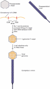
During the in vivo assembly of λ virions within infected host cells, viral heads and tails initially are assembled separately, from multiple copies of the various proteins that compose these complex structures. Replication of λ DNA in a host cell generates long multimeric DNA molecules, called concatomers, that consist of multiple copies of the viral genome linked end to end and separated by specific nucleotide sequences called COS sites. Two λ proteins, designated Nu1 and A, bind to COS sites and direct insertion of the DNA lying between two adjacent COS sites into a preassembled head. This process results in the packaging of a single ≈50-kb λ genome from the multimeric concatomer into each preassembled head. Host-cell chromosomal DNA is not inserted into the λ heads because it does not contain any copies of the COS sequence. Once λ DNA is inserted into a preassembled λ head, the preassembled tail is attached, producing a complete virion.
Bacteriophage λ Can Be Modified for Use as a Cloning Vector and Assembled in Vitro
Bacteriophage λ is probably the most extensively studied bacterial virus, and a great deal is known about its molecular biology and genetics. A λ phage virion has a head, which contains the viral DNA genome, and a tail, which functions in infecting E. coli host cells. When λ DNA enters the host-cell cytoplasm following infection, it undergoes either lytic or lysogenic growth. In lytic growth, the viral DNA is replicated and assembled into more than 100 progeny virions in each infected cell, killing the cell in the process and releasing the replicated virions. In lysogenic growth, the viral DNA inserts into the bacterial chromosome, where it is passively replicated along with the host-cell chromosome as the cell grows and divides.
The bacteriophage genome. (a) Electron micrograph of bacteriophage λ virion. The genome is contained within the head. (b) Simplified map of the λ phage genome.
The λ genes encoding the head and tail proteins as well as various proteins involved in the lytic and lysogenic growth pathways are clustered in discrete regions of the ≈50-kb viral genome. When bacteriophage λ is used as a cloning vector, it must be capable of lytic growth, but other viral functions are irrelevant. Consequently, the genes involved in the lysogenic pathway and other viral genes not essential for the lytic pathway are removed from the viral DNA and replaced with the DNA to be cloned. Up to ≈25 kb of foreign DNA can be inserted into the λ genome, resulting in a recombinant DNA that can be packaged in vitro to form virions capable of replicating and forming plaques on E. coli host cells.
During the in vivo assembly of λ virions within infected host cells, viral heads and tails initially are assembled separately, from multiple copies of the various proteins that compose these complex structures. Replication of λ DNA in a host cell generates long multimeric DNA molecules, called concatomers, that consist of multiple copies of the viral genome linked end to end and separated by specific nucleotide sequences called COS sites. Two λ proteins, designated Nu1 and A, bind to COS sites and direct insertion of the DNA lying between two adjacent COS sites into a preassembled head. This process results in the packaging of a single ≈50-kb λ genome from the multimeric concatomer into each preassembled head. Host-cell chromosomal DNA is not inserted into the λ heads because it does not contain any copies of the COS sequence. Once λ DNA is inserted into a preassembled λ head, the preassembled tail is attached, producing a complete virion.
Friday, April 23, 2010
News of Human Genome Sciences get positive results from phase-3 trial of Benlysta in SLE
At Week 76 in the BLISS-76 study, belimumab plus standard of care showed higher response rates compared with placebo plus standard of care as measured by the SLE Responder Index; however, this secondary endpoint did not reach statistical significance. Study results also showed that belimumab continued to be generally well tolerated, as demonstrated by a similar rate of discontinuations due to adverse events across treatment groups, with overall adverse event rates comparable between belimumab and placebo treatment groups.
“A positive overall picture has emerged from our pivotal phase-3 studies of Benlysta, including its achievement of statistical significance on the primary efficacy endpoint at Week 52 with a favourable safety profile in both BLISS-52 and BLISS-76,” said H Thomas Watkins, president and chief executive officer, HGS. “We view the results of these studies as strongly supportive of our view that Benlysta has the potential to become the first new approved drug in more than 50 years for people living with systemic lupus.”
Carlo Russo, senior vice president, Biopharm Development, GSK, said, “Based on the totality of data in BLISS-52 and BLISS-76, we believe that belimumab could deliver a significant therapeutic option for patients with lupus, a chronic condition which has a devastating effect on the lives of patients living with the disease.”
The data from the BLISS-76 study were previously analysed after 52 weeks in accord with the study protocol, in support of a potential Biologics License Application in the United States and Marketing Authorization Applications in Europe and other regions. The primary efficacy endpoints in both pivotal phase-3 studies of belimumab, BLISS-52 and BLISS-76, were the patient response rates at Week 52 as measured by the SLE Responder Index. BLISS-76 then continued for an additional 24 weeks. Belimumab is an investigational drug and the first in a new class of drugs called BLyS-specific inhibitors. Belimumab is being developed by HGS and GSK under a co-development and commercialisation agreement entered into in 2006.
“These new data from BLISS-76 provide additional evidence of the beneficial effect of belimumab despite not reaching statistical significance on the secondary endpoint. The results of our phase-3 trials support a potentially important role for belimumab added to standard of care for the treatment of seropositive patients with systemic lupus,” said David C. Stump, executive vice president, Research and Development, HGS. “We and GSK are working together to complete and submit regulatory applications for belimumab in the United States and Europe in the second quarter of this year. We look forward to the full presentation of BLISS-76 52-week and 76-week results at appropriate scientific meetings later this year.”
Based on an intention-to-treat (ITT) analysis, patient response rates for belimumab plus standard of care versus placebo plus standard of care, as measured by the SLE Responder Index (SRI) at Week 76, were: 38.5 per cent for 10 mg/kg belimumab, 39.1 per cent for 1 mg/kg belimumab, and 32.4 per cent for placebo (p=0.13 and p=0.11 for 10 mg/kg and 1 mg/kg belimumab, respectively vs. placebo).
“A positive overall picture has emerged from our pivotal phase-3 studies of Benlysta, including its achievement of statistical significance on the primary efficacy endpoint at Week 52 with a favourable safety profile in both BLISS-52 and BLISS-76,” said H Thomas Watkins, president and chief executive officer, HGS. “We view the results of these studies as strongly supportive of our view that Benlysta has the potential to become the first new approved drug in more than 50 years for people living with systemic lupus.”
Carlo Russo, senior vice president, Biopharm Development, GSK, said, “Based on the totality of data in BLISS-52 and BLISS-76, we believe that belimumab could deliver a significant therapeutic option for patients with lupus, a chronic condition which has a devastating effect on the lives of patients living with the disease.”
The data from the BLISS-76 study were previously analysed after 52 weeks in accord with the study protocol, in support of a potential Biologics License Application in the United States and Marketing Authorization Applications in Europe and other regions. The primary efficacy endpoints in both pivotal phase-3 studies of belimumab, BLISS-52 and BLISS-76, were the patient response rates at Week 52 as measured by the SLE Responder Index. BLISS-76 then continued for an additional 24 weeks. Belimumab is an investigational drug and the first in a new class of drugs called BLyS-specific inhibitors. Belimumab is being developed by HGS and GSK under a co-development and commercialisation agreement entered into in 2006.
“These new data from BLISS-76 provide additional evidence of the beneficial effect of belimumab despite not reaching statistical significance on the secondary endpoint. The results of our phase-3 trials support a potentially important role for belimumab added to standard of care for the treatment of seropositive patients with systemic lupus,” said David C. Stump, executive vice president, Research and Development, HGS. “We and GSK are working together to complete and submit regulatory applications for belimumab in the United States and Europe in the second quarter of this year. We look forward to the full presentation of BLISS-76 52-week and 76-week results at appropriate scientific meetings later this year.”
Based on an intention-to-treat (ITT) analysis, patient response rates for belimumab plus standard of care versus placebo plus standard of care, as measured by the SLE Responder Index (SRI) at Week 76, were: 38.5 per cent for 10 mg/kg belimumab, 39.1 per cent for 1 mg/kg belimumab, and 32.4 per cent for placebo (p=0.13 and p=0.11 for 10 mg/kg and 1 mg/kg belimumab, respectively vs. placebo).
The Genome types and C Value
The Genome:
A genome is the complete collection of hereditary information for an individual organism. In cellular life forms, the hereditary information exists as DNA. There are two fundamentally distinct types of cells in the living world, prokaryotic and eukaryotic, and the organization of genomes differs in these two types of cells.
Types:
Most biological entities that are more complex than a virus sometimes or always carry additional genetic material besides that which resides in their chromosomes. In some contexts, such as sequencing the genome of a pathogenic microbe, "genome" is meant to include information stored on this auxiliary material, which is carried in plasmids. In such circumstances then, "genome" describes all of the genes and information on non-coding DNA that have the potential to be present.
In eukaryotes such as plants, protozoa and animals, however, "genome" carries the typical connotation of only information on chromosomal DNA. So although these organisms contain chloroplasts and/or mitochondria that have their own DNA, the genetic information contained by DNA within these organelles is not considered part of the genome. In fact, mitochondria are sometimes said to have their own genome often referred to as the "mitochondrial genome". The DNA found within the chloroplast may be referred to as the "plastome".
Genome Size or C Value:
The C value is the amount of DNA in a haploid complement. Currently, the amount is reported as the total number of base pairs. Generally, more complex organisms have more DNA. For example, the haploid complement of Homo sapiens DNA contains between 3.12 and 3.2 gigabases (the prefix "giga" denotes billions), while the haploid complement of yeast (Saccharomyces cerevisiae) DNA contains 12,057,500 base pairs.
Unexpected genomic sizes occur, however, in a condition called the C value paradox. Two closely related species can have widely divergent amounts of DNA. For example, Paramecium caudatum has a C value of 8,600,000 kilobases (where the prefix "kilo" denotes thousands) while its near relative P. aurelia has a C value of just 190,000 kilobases. Another paradoxical circumstance occurs when a simpler organism has a C value higher than a more complex organism. For example, Amphiuma means (a newt) and Amoeba dubia (an amoeba) have, respectively, C values that are 26 and 209 times the C value of humans.
A genome is the complete collection of hereditary information for an individual organism. In cellular life forms, the hereditary information exists as DNA. There are two fundamentally distinct types of cells in the living world, prokaryotic and eukaryotic, and the organization of genomes differs in these two types of cells.
Types:
Most biological entities that are more complex than a virus sometimes or always carry additional genetic material besides that which resides in their chromosomes. In some contexts, such as sequencing the genome of a pathogenic microbe, "genome" is meant to include information stored on this auxiliary material, which is carried in plasmids. In such circumstances then, "genome" describes all of the genes and information on non-coding DNA that have the potential to be present.
In eukaryotes such as plants, protozoa and animals, however, "genome" carries the typical connotation of only information on chromosomal DNA. So although these organisms contain chloroplasts and/or mitochondria that have their own DNA, the genetic information contained by DNA within these organelles is not considered part of the genome. In fact, mitochondria are sometimes said to have their own genome often referred to as the "mitochondrial genome". The DNA found within the chloroplast may be referred to as the "plastome".
Genome Size or C Value:
The C value is the amount of DNA in a haploid complement. Currently, the amount is reported as the total number of base pairs. Generally, more complex organisms have more DNA. For example, the haploid complement of Homo sapiens DNA contains between 3.12 and 3.2 gigabases (the prefix "giga" denotes billions), while the haploid complement of yeast (Saccharomyces cerevisiae) DNA contains 12,057,500 base pairs.
Unexpected genomic sizes occur, however, in a condition called the C value paradox. Two closely related species can have widely divergent amounts of DNA. For example, Paramecium caudatum has a C value of 8,600,000 kilobases (where the prefix "kilo" denotes thousands) while its near relative P. aurelia has a C value of just 190,000 kilobases. Another paradoxical circumstance occurs when a simpler organism has a C value higher than a more complex organism. For example, Amphiuma means (a newt) and Amoeba dubia (an amoeba) have, respectively, C values that are 26 and 209 times the C value of humans.
Sunday, April 11, 2010
change-your-biochemistry-to-get-bigger penis
Are you unhappy with the size of your manhood and would desperately like to know what you can do about it? If so, you've come to the right place because I can help you to add inches to your penis in a matter of weeks. There's nothing to buy, so it wont cost you a penny and the whole process is exactly the same one that your body followed during puberty, meaning that it is 100% safe too. So, how do you make your manhood grow? The answer is simple - you just have to use natural enlargement...
Is natural enlargement a tried and tested approach? Does it really work?
The answer to both of these questions is yes - I have personally added 4 inches to my penis this way! Also, it has been proven to work by various scientists all around the world, and this makes it truely unique. I would never have believed that getting a larger member could be done so effortlessly, but with natural enlargement it couldn't have been simpler! The key is to get your body to do the work - once you do that, natural growth becomes much easier.
What is the process that it follows?
Like I've already mentioned, to grow, you need to follow the same process that caused a change in your size during puberty. And that means, you will need to put back any biochemicals that were originally around at that time. By changing your biochemistry, you can unlock your body's true potential to grow - and the results are amazing.
Is there anything you can do to grow even faster?
The best thing to accelerate growth is exercise. By using a natural enlargement scheme, you will learn how to change your biochemistry and how to exercise - and these are the two key parts to your success. This is your way to finally get the manhood you deserve - you'd be fool to miss out!
After only 4 weeks, I doubled in size and I have to say it feels great! I'm more confident and my life is on the up - you could feel this way too.
Is natural enlargement a tried and tested approach? Does it really work?
The answer to both of these questions is yes - I have personally added 4 inches to my penis this way! Also, it has been proven to work by various scientists all around the world, and this makes it truely unique. I would never have believed that getting a larger member could be done so effortlessly, but with natural enlargement it couldn't have been simpler! The key is to get your body to do the work - once you do that, natural growth becomes much easier.
What is the process that it follows?
Like I've already mentioned, to grow, you need to follow the same process that caused a change in your size during puberty. And that means, you will need to put back any biochemicals that were originally around at that time. By changing your biochemistry, you can unlock your body's true potential to grow - and the results are amazing.
Is there anything you can do to grow even faster?
The best thing to accelerate growth is exercise. By using a natural enlargement scheme, you will learn how to change your biochemistry and how to exercise - and these are the two key parts to your success. This is your way to finally get the manhood you deserve - you'd be fool to miss out!
After only 4 weeks, I doubled in size and I have to say it feels great! I'm more confident and my life is on the up - you could feel this way too.
How to Study Biochemistry
Biochemistry is a notorious course for demanding a high-volume of information in a short amount of time. However, there are studying methods to assist students in learning efficiently and effectively. I have studied and interviewed groups of medical and science students that have mastered their course work. It is true that there are specific and detailed guidelines that these students adhere to and credit for their academic success. The successful student must excel in visualizing relationships, memorizing facts, and reciting complex metabolic reactions of the human body. With some time and applying these strategies and tips from past honor students of Biochemistry, you will greatly improve your academic performance.Study Skill #1 - Do NOT procrastinate. The most obvious, and yet least followed advice by students. Biochemistry is a high-volume course that progresses and builds its concepts on the fundamentals. Moreover, many pathways and reactions require memorization and must be acquired over time. The last thing you want to do is cram for this course.Study Skill #2 - Know the terminology and nomenclature, it will make things much easier down the road. An enzyme or protein will often have its function built into its name. Take Protein Kinase A for example. As a member of the Kinases, it will almost always add a phosphate group to its substrate. Or, take Alcohol Dehydrogenase, structures that are Dehydrogenases always oxidize a substrate. In this case, it oxidizes alcohols into aldehydes and ketones. Once you get this down, you will begin to recognize names and automatically correlate them with a specific function.Study Skill #3 - Start with the big picture. There is no doubt that you will have to memorize multi-step metabolic pathways. The best way to do this is to start with the easy steps and understand the overall flow of the reaction. First, write only the substrates and products in order. Do this repeatedly, until it is memorized. Then add the enzymes. Then continue to add co-factors and by-products. If necessary, label each as an exer- or endergonic reaction. Use the nomenclature to help you remember what is going on in each step. For example, Phosphofructokinase-1 - adds a phosphate group (phospho-kinase) to the molecule fructose (-fructo-) at the first position (-1). By breaking down the pathways and focusing on the terminology it will greatly speed up your ability to memorize them.Study Skill #4 - Buy a dry erase board. Use this to memorize the pathways and any other reactions you have to know. There are no short-cuts, but writing things out reinforces them in your memory. It tends to be much more efficient than staring and reciting from your textbook.Study Skill #5 - Know the purpose of a reaction. Take the Bohr Effect for example. An increase in (decrease in pH), temperature, and 2,3-BPG all occur in active skeletal muscle. They also all encourage release from hemoglobin. This makes sense if you think that working muscle is metabolic tissue and needs oxygen to survive. Incorporating the larger concept will also allow you to predict the flow of reactions in other situations throughout the body.Study Skill #6 - Stare at the graphs and plots. These questions are virtually freebies on exams because all the information you need to solve them is included. Know what the x- and y-intercept, the slope, and the area under the graph represent. Know what makes the graphed line move to the right or left. You will absolutely be asked about the Michaelis-Menten graph and the Hemoglobin dissociation curve - these are staples of biochemistry.Study Skill #7 - Seek to understand first, and then memorize. Like many other courses, biochemistry can be overwhelming at first. There is no easy way to memorize every amino acid or metabolic reaction. But students always claim that if they take the time to first get the concept down, the memorizing is not as difficult as it once seemed. Stay focused, break it down into small steps, and practice.Jordan Castle is a medical student in Detroit, Michigan. His work spans many different aspects of the learning process and aims to help students excel in their individual courses.
Saturday, March 27, 2010
A Shifting Drug Industry Means New Opportunities in Translational Research
New opportunities
Despite all the changes, pharmaceutical companies are maintaining a strong internal development program in areas with large markets such as oncology, neuroscience, and diabetes/obesity; and they are hiring people whose skills fit with their drug-development programs. Furthermore, the shifts in the industry may herald a new, more fluid division of labor where nontraditional partnerships take on the earliest stages of drug development. "I think it may be up to smaller biotech companies and academia to come up with new drugs," Littman says. "And smaller biotech companies are very interested in generating biomarkers, working on proof of concept, and testing in smaller patient populations."
It's a modular approach to drug discovery and translational research that both FitzGerald and others believe will become prevalent. They also think this will necessitate changes in the way industry and academia handle things such as intellectual property. People choosing industry careers should be prepared and adaptable.
"I think there is a lot of uncertainty out there in the world right now," says Will West, chief executive at CellCentric, a biotechnology company in Cambridge, United Kingdom. "If you are a young graduate, I can see how basing a career in industry may seem like a difficult choice to make. I see the opposite. I think in a changing model, there is opportunity for bright people to take advantage of that change and be drivers for the solutions.
Despite all the changes, pharmaceutical companies are maintaining a strong internal development program in areas with large markets such as oncology, neuroscience, and diabetes/obesity; and they are hiring people whose skills fit with their drug-development programs. Furthermore, the shifts in the industry may herald a new, more fluid division of labor where nontraditional partnerships take on the earliest stages of drug development. "I think it may be up to smaller biotech companies and academia to come up with new drugs," Littman says. "And smaller biotech companies are very interested in generating biomarkers, working on proof of concept, and testing in smaller patient populations."
It's a modular approach to drug discovery and translational research that both FitzGerald and others believe will become prevalent. They also think this will necessitate changes in the way industry and academia handle things such as intellectual property. People choosing industry careers should be prepared and adaptable.
"I think there is a lot of uncertainty out there in the world right now," says Will West, chief executive at CellCentric, a biotechnology company in Cambridge, United Kingdom. "If you are a young graduate, I can see how basing a career in industry may seem like a difficult choice to make. I see the opposite. I think in a changing model, there is opportunity for bright people to take advantage of that change and be drivers for the solutions.
Sunday, March 7, 2010
Recombinant DNA Technology Has Revolutionized All Aspects of Biology
recombinant DNA technology, which has permitted biology to move from an exclusively analytical science to a
synthetic one. New combinations of unrelated genes can be constructed in the laboratory by applying recombinant DNA
techniques. These novel combinations can be cloned amplified manyfold by introducing them into suitable cells,
where they are replicated by the DNA-synthesizing machinery of the host. The inserted genes are often transcribed and
translated in their new setting. What is most striking is that the genetic endowment of the host can be permanently
altered in a designed way.
Restriction Enzymes and DNA Ligase Are Key Tools in Forming Recombinant
DNA Molecules
Let us begin by seeing how novel DNA molecules can be constructed in the laboratory. A DNA fragment of interest is
covalently joined to a DNA vector. The essential feature of a vector is that it can replicate autonomously in an
appropriate host. Plasmids (naturally occurring circles of DNA that act as accessory chromosomes in bacteria) and
bacteriophage l , a virus, are choice vectors for cloning in E. coli. The vector can be prepared for accepting a new DNA
fragment by cleaving it at a single specific site with a restriction enzyme. For example, the plasmid pSC101, a 9.9-kb
double-helical circular DNA molecule, is split at a unique site by the EcoRI restriction enzyme. The staggered cuts made
by this enzyme produce complementary single-stranded ends, which have specific affinity for each other and hence are
known as cohesive or sticky ends. Any DNA fragment can be inserted into this plasmid if it has the same cohesive ends.
Such a fragment can be prepared from a larger piece of DNA by using the same restriction enzyme as was used to open
the plasmid DNA
The single-stranded ends of the fragment are then complementary to those of the cut plasmid. The DNA fragment and
the cut plasmid can be annealed and then joined by DNA ligase, which catalyzes the formation of a phosphodiester bond
at a break in a DNA chain. DNA ligase requires a free 3 -hydroxyl group and a 5 -phosphoryl group. Furthermore, the
chains joined by ligase must be in a double helix. An energy source such as ATP or NAD+ is required for the joining
reaction,
This cohesive-end method for joining DNA molecules can be made general by using a short, chemically synthesized
DNA linker that can be cleaved by restriction enzymes. First, the linker is covalently joined to the ends of a DNA
fragment or vector. For example, the 5 ends of a decameric linker and a DNA molecule are phosphorylated by
polynucleotide kinase and then joined by the ligase from T4 phage . This ligase can form a covalent bond
between blunt-ended (flush-ended) double-helical DNA molecules. Cohesive ends are produced when these terminal
extensions are cut by an appropriate restriction enzyme. Thus, cohesive ends corresponding to a particular restriction
enzyme can be added to virtually any DNA molecule. We see here the fruits of combining enzymatic and synthetic chemical approaches in crafting new DNA molecules.
synthetic one. New combinations of unrelated genes can be constructed in the laboratory by applying recombinant DNA
techniques. These novel combinations can be cloned amplified manyfold by introducing them into suitable cells,
where they are replicated by the DNA-synthesizing machinery of the host. The inserted genes are often transcribed and
translated in their new setting. What is most striking is that the genetic endowment of the host can be permanently
altered in a designed way.
Restriction Enzymes and DNA Ligase Are Key Tools in Forming Recombinant
DNA Molecules
Let us begin by seeing how novel DNA molecules can be constructed in the laboratory. A DNA fragment of interest is
covalently joined to a DNA vector. The essential feature of a vector is that it can replicate autonomously in an
appropriate host. Plasmids (naturally occurring circles of DNA that act as accessory chromosomes in bacteria) and
bacteriophage l , a virus, are choice vectors for cloning in E. coli. The vector can be prepared for accepting a new DNA
fragment by cleaving it at a single specific site with a restriction enzyme. For example, the plasmid pSC101, a 9.9-kb
double-helical circular DNA molecule, is split at a unique site by the EcoRI restriction enzyme. The staggered cuts made
by this enzyme produce complementary single-stranded ends, which have specific affinity for each other and hence are
known as cohesive or sticky ends. Any DNA fragment can be inserted into this plasmid if it has the same cohesive ends.
Such a fragment can be prepared from a larger piece of DNA by using the same restriction enzyme as was used to open
the plasmid DNA
The single-stranded ends of the fragment are then complementary to those of the cut plasmid. The DNA fragment and
the cut plasmid can be annealed and then joined by DNA ligase, which catalyzes the formation of a phosphodiester bond
at a break in a DNA chain. DNA ligase requires a free 3 -hydroxyl group and a 5 -phosphoryl group. Furthermore, the
chains joined by ligase must be in a double helix. An energy source such as ATP or NAD+ is required for the joining
reaction,
This cohesive-end method for joining DNA molecules can be made general by using a short, chemically synthesized
DNA linker that can be cleaved by restriction enzymes. First, the linker is covalently joined to the ends of a DNA
fragment or vector. For example, the 5 ends of a decameric linker and a DNA molecule are phosphorylated by
polynucleotide kinase and then joined by the ligase from T4 phage . This ligase can form a covalent bond
between blunt-ended (flush-ended) double-helical DNA molecules. Cohesive ends are produced when these terminal
extensions are cut by an appropriate restriction enzyme. Thus, cohesive ends corresponding to a particular restriction
enzyme can be added to virtually any DNA molecule. We see here the fruits of combining enzymatic and synthetic chemical approaches in crafting new DNA molecules.
PCR Is a Powerful Technique in Medical Diagnostics, Forensics, and Molecular
In 1984, Kary Mullis devised an ingenious method called the polymerase chain reaction (PCR) for amplifying specific
DNA sequences. Consider a DNA duplex consisting of a target sequence surrounded by nontarget DNA. Millions of the
target sequences can be readily obtained by PCR if the flanking sequences of the target are known. PCR is carried out by
adding the following components to a solution containing the target sequence: (1) a pair of primers that hybridize with
the flanking sequences of the target, (2) all four deoxyribonucleoside triphosphates (dNTPs), and (3) a heat-stable DNA
polymerase. A PCR cycle consists of three steps (Figure 6.8).
1. Strand separation. The two strands of the parent DNA molecule are separated by heating the solution to 95°C for 15 s.
2. Hybridization of primers. The solution is then abruptly cooled to 54°C to allow each primer to hybridize to a DNA
strand. One primer hybridizes to the 3 -end of the target on one strand, and the other primer hybridizes to the 3 end on
the complementary target strand. Parent DNA duplexes do not form, because the primers are present in large excess.
Primers are typically from 20 to 30 nucleotides long.
3. DNA synthesis. The solution is then heated to 72°C, the optimal temperature for Taq DNA polymerase. This heatstable
polymerase comes from T hermus aq uaticus, a thermophilic bacterium that lives in hot springs. The polymerase
elongates both primers in the direction of the target sequence because DNA synthesis is in the 5 -to-3 direction. DNA
synthesis takes place on both strands but extends beyond the target sequence.
These three steps strand separation, hybridization of primers, and DNA synthesis constitute one cycle of the PCR
amplification and can be carried out repetitively just by changing the temperature of the reaction mixture. The
thermostability of the polymerase makes it feasible to carry out PCR in a closed container; no reagents are added after
the first cycle. The duplexes are heated to begin the second cycle, which produces four duplexes, and then the third cycle
is initiated . At the end of the third cycle, two short strands appear that constitute only the target
sequence the sequence including and bounded by the primers. Subsequent cycles will amplify the target sequence
exponentially. The larger strands increase in number arithmetically and serve as a source for the synthesis of more short
strands. Ideally, after n cycles, this sequence is amplified 2 n -fold. The amplification is a millionfold after 20 cycles and
a billionfold after 30 cycles, which can be carried out in less than an hour.
Several features of this remarkable method for amplifying DNA are noteworthy. First, the sequence of the target need
not be known. All that is required is knowledge of the flanking sequences. Second, the target can be much larger than the
primers. Targets larger than 10 kb have been amplified by PCR. Third, primers do not have to be perfectly matched to
flanking sequences to amplify targets. With the use of primers derived from a gene of known sequence, it is possible to
search for variations on the theme. In this way, families of genes are being discovered by PCR. Fourth, PCR is highly
specific because of the stringency of hybridization at high temperature (54°C). Stringency is the required closeness of the
match between primer and target, which can be controlled by temperature and salt. At high temperatures, the only DNA
that is amplified is that situated between primers that have hybridized. A gene constituting less than a millionth of the
total DNA of a higher organism is accessible by PCR. Fifth, PCR is exquisitely sensitive. A single DNA molecule can be
amplified and detected.
PCR can provide valuable diagnostic information in medicine. Bacteria and viruses can be readily detected with the use
of specific primers. For example, PCR can reveal the presence of human immunodeficiency virus in people who have
not mounted an immune response to this pathogen and would therefore be missed with an antibody assay. Finding
Mycobacterium tuberculosis bacilli in tissue specimens is slow and laborious. With PCR, as few as 10 tubercle bacilli
per million human cells can be readily detected. PCR is a promising method for the early detection of certain cancers.
This technique can identify mutations of certain growth-control genes, such as the ras genes (Section 15.4.2). The
capacity to greatly amplify selected regions of DNA can also be highly informative in monitoring cancer chemotherapy.
Tests using PCR can detect when cancerous cells have been eliminated and treatment can be stopped; they can also
detect a relapse and the need to immediately resume treatment. PCR is ideal for detecting leukemias caused by
chromosomal rearrangements.
PCR is also having an effect in forensics and legal medicine. An individual DNA profile is highly distinctive because
many genetic loci are highly variable within a population. For example, variations at a specific one of these locations
determines a person's HLA type (human leukocyte antigen type); organ transplants are rejected when the HLA types of
the donor and recipient are not sufficiently matched. PCR amplification of multiple genes is being used to establish
biological parentage in disputed paternity and immigration cases. Analyses of blood stains and semen samples by PCR
have implicated guilt or innocence in numerous assault and rape cases. The root of a single shed hair found at a crime
scene contains enough DNA for typing by PCR
DNA is a remarkably stable molecule, particularly when relatively shielded from air, light, and water. Under such
circumstances, large fragments of DNA can remain intact for thousands of years or longer. PCR provides an ideal
method for amplifying such ancient DNA molecules so that they can be detected and characterized (Section 7.5.1). PCR
can also be used to amplify DNA from microorganisms that have not yet been isolated and cultured. As will be discussed
in the next chapter, sequences from these PCR products can be sources of considerable insight into evolutionary
relationships between organisms.
DNA sequences. Consider a DNA duplex consisting of a target sequence surrounded by nontarget DNA. Millions of the
target sequences can be readily obtained by PCR if the flanking sequences of the target are known. PCR is carried out by
adding the following components to a solution containing the target sequence: (1) a pair of primers that hybridize with
the flanking sequences of the target, (2) all four deoxyribonucleoside triphosphates (dNTPs), and (3) a heat-stable DNA
polymerase. A PCR cycle consists of three steps (Figure 6.8).
1. Strand separation. The two strands of the parent DNA molecule are separated by heating the solution to 95°C for 15 s.
2. Hybridization of primers. The solution is then abruptly cooled to 54°C to allow each primer to hybridize to a DNA
strand. One primer hybridizes to the 3 -end of the target on one strand, and the other primer hybridizes to the 3 end on
the complementary target strand. Parent DNA duplexes do not form, because the primers are present in large excess.
Primers are typically from 20 to 30 nucleotides long.
3. DNA synthesis. The solution is then heated to 72°C, the optimal temperature for Taq DNA polymerase. This heatstable
polymerase comes from T hermus aq uaticus, a thermophilic bacterium that lives in hot springs. The polymerase
elongates both primers in the direction of the target sequence because DNA synthesis is in the 5 -to-3 direction. DNA
synthesis takes place on both strands but extends beyond the target sequence.
These three steps strand separation, hybridization of primers, and DNA synthesis constitute one cycle of the PCR
amplification and can be carried out repetitively just by changing the temperature of the reaction mixture. The
thermostability of the polymerase makes it feasible to carry out PCR in a closed container; no reagents are added after
the first cycle. The duplexes are heated to begin the second cycle, which produces four duplexes, and then the third cycle
is initiated . At the end of the third cycle, two short strands appear that constitute only the target
sequence the sequence including and bounded by the primers. Subsequent cycles will amplify the target sequence
exponentially. The larger strands increase in number arithmetically and serve as a source for the synthesis of more short
strands. Ideally, after n cycles, this sequence is amplified 2 n -fold. The amplification is a millionfold after 20 cycles and
a billionfold after 30 cycles, which can be carried out in less than an hour.
Several features of this remarkable method for amplifying DNA are noteworthy. First, the sequence of the target need
not be known. All that is required is knowledge of the flanking sequences. Second, the target can be much larger than the
primers. Targets larger than 10 kb have been amplified by PCR. Third, primers do not have to be perfectly matched to
flanking sequences to amplify targets. With the use of primers derived from a gene of known sequence, it is possible to
search for variations on the theme. In this way, families of genes are being discovered by PCR. Fourth, PCR is highly
specific because of the stringency of hybridization at high temperature (54°C). Stringency is the required closeness of the
match between primer and target, which can be controlled by temperature and salt. At high temperatures, the only DNA
that is amplified is that situated between primers that have hybridized. A gene constituting less than a millionth of the
total DNA of a higher organism is accessible by PCR. Fifth, PCR is exquisitely sensitive. A single DNA molecule can be
amplified and detected.
PCR can provide valuable diagnostic information in medicine. Bacteria and viruses can be readily detected with the use
of specific primers. For example, PCR can reveal the presence of human immunodeficiency virus in people who have
not mounted an immune response to this pathogen and would therefore be missed with an antibody assay. Finding
Mycobacterium tuberculosis bacilli in tissue specimens is slow and laborious. With PCR, as few as 10 tubercle bacilli
per million human cells can be readily detected. PCR is a promising method for the early detection of certain cancers.
This technique can identify mutations of certain growth-control genes, such as the ras genes (Section 15.4.2). The
capacity to greatly amplify selected regions of DNA can also be highly informative in monitoring cancer chemotherapy.
Tests using PCR can detect when cancerous cells have been eliminated and treatment can be stopped; they can also
detect a relapse and the need to immediately resume treatment. PCR is ideal for detecting leukemias caused by
chromosomal rearrangements.
PCR is also having an effect in forensics and legal medicine. An individual DNA profile is highly distinctive because
many genetic loci are highly variable within a population. For example, variations at a specific one of these locations
determines a person's HLA type (human leukocyte antigen type); organ transplants are rejected when the HLA types of
the donor and recipient are not sufficiently matched. PCR amplification of multiple genes is being used to establish
biological parentage in disputed paternity and immigration cases. Analyses of blood stains and semen samples by PCR
have implicated guilt or innocence in numerous assault and rape cases. The root of a single shed hair found at a crime
scene contains enough DNA for typing by PCR
DNA is a remarkably stable molecule, particularly when relatively shielded from air, light, and water. Under such
circumstances, large fragments of DNA can remain intact for thousands of years or longer. PCR provides an ideal
method for amplifying such ancient DNA molecules so that they can be detected and characterized (Section 7.5.1). PCR
can also be used to amplify DNA from microorganisms that have not yet been isolated and cultured. As will be discussed
in the next chapter, sequences from these PCR products can be sources of considerable insight into evolutionary
relationships between organisms.
Exploring Genes
Recombinant DNA technology has revolutionized biochemistry since it came into being in the 1970s. The genetic
endowment of organisms can now be precisely changed in designed ways. Recombinant DNA technology is a fruit of
several decades of basic research on DNA, RNA, and viruses. It depends, first, on having enzymes that can cut, join, and
replicate DNA and reverse transcribe RNA. Restriction enzymes cut very long DNA molecules into specific fragments
that can be manipulated; DNA ligases join the fragments together. The availability of many kinds of restriction enzymes
and DNA ligases makes it feasible to treat DNA sequences as modules that can be moved at will from one DNA
molecule to another. Thus, recombinant DNA technology is based on nucleic acid enzymology.
A second foundation is the base-pairing language that allows complementary sequences to recognize and bind to each
other. Hybridization with complementary DNA or RNA probes is a sensitive and powerful means of detecting specific
nucleotide sequences. In recombinant DNA technology, base-pairing is used to construct new combinations of DNA as
well as to detect and amplify particular sequences. This revolutionary technology is also critically dependent on our
understanding of viruses, the ultimate parasites. Viruses efficiently deliver their own DNA (or RNA) into hosts,
subverting them either to replicate the viral genome and produce viral proteins or to incorporate viral DNA into the host
genome. Likewise, plasmids, which are accessory chromosomes found in bacteria, have been indispensable in
recombinant DNA technology.
These new methods have wide-ranging benefits. Entire genomes, including the human genome, are being deciphered.
New insights are emerging, for example, into the regulation of gene expression in cancer and development and the
evolutionary history of proteins as well as organisms. New proteins can be created by altering genes in specific ways to
provide detailed views into protein function. Clinically useful proteins, such as hormones, are now synthesized by
recombinant DNA techniques. Crops are being generated to resist pests and harsh conditions. The new opportunities
opened by recombinant DNA technology promise to have broad effects.
The Basic Tools of Gene Exploration
The rapid progress in biotechnology indeed its very existence is a result of a relatively few techniques.
1. Restriction-enzyme analysis. Restriction enzymes are precise, molecular scalpels that allow the investigator to
manipulate DNA segments.
2. Blotting techniques. The Southern and Northern blots are used to separate and characterize DNA and RNA,
respectively. The Western blot, which uses antibodies to characterize proteins, was described in Section 4.3.4.
3. DNA sequencing. The precise nucleotide sequence of a molecule of DNA can be determined. Sequencing has yielded a
wealth of information concerning gene architecture, the control of gene expression, and protein structure.
4. Solid-phase synthesis of nucleic acids. Precise sequences of nucleic acids can be synthesized de novo and used to
identify or amplify other nucleic acids.
5. The polymerase chain reaction (PCR). The polymerase chain reaction leads to a billionfold amplification of a segment
of DNA. One molecule of DNA can be amplified to quantities that permit characterization and manipulation. This
powerful technique is being used to detect pathogens and genetic diseases, to determine the source of a hair left at the
scene of a crime, and to resurrect genes from fossils.
A final tool, the use of which will be highlighted in the next chapter, is the computer. Without the computer, it would be
impossible to catalog, access, and characterize the abundant information, especially DNA sequence information, that the techniques just outlined are rapidly generating.
endowment of organisms can now be precisely changed in designed ways. Recombinant DNA technology is a fruit of
several decades of basic research on DNA, RNA, and viruses. It depends, first, on having enzymes that can cut, join, and
replicate DNA and reverse transcribe RNA. Restriction enzymes cut very long DNA molecules into specific fragments
that can be manipulated; DNA ligases join the fragments together. The availability of many kinds of restriction enzymes
and DNA ligases makes it feasible to treat DNA sequences as modules that can be moved at will from one DNA
molecule to another. Thus, recombinant DNA technology is based on nucleic acid enzymology.
A second foundation is the base-pairing language that allows complementary sequences to recognize and bind to each
other. Hybridization with complementary DNA or RNA probes is a sensitive and powerful means of detecting specific
nucleotide sequences. In recombinant DNA technology, base-pairing is used to construct new combinations of DNA as
well as to detect and amplify particular sequences. This revolutionary technology is also critically dependent on our
understanding of viruses, the ultimate parasites. Viruses efficiently deliver their own DNA (or RNA) into hosts,
subverting them either to replicate the viral genome and produce viral proteins or to incorporate viral DNA into the host
genome. Likewise, plasmids, which are accessory chromosomes found in bacteria, have been indispensable in
recombinant DNA technology.
These new methods have wide-ranging benefits. Entire genomes, including the human genome, are being deciphered.
New insights are emerging, for example, into the regulation of gene expression in cancer and development and the
evolutionary history of proteins as well as organisms. New proteins can be created by altering genes in specific ways to
provide detailed views into protein function. Clinically useful proteins, such as hormones, are now synthesized by
recombinant DNA techniques. Crops are being generated to resist pests and harsh conditions. The new opportunities
opened by recombinant DNA technology promise to have broad effects.
The Basic Tools of Gene Exploration
The rapid progress in biotechnology indeed its very existence is a result of a relatively few techniques.
1. Restriction-enzyme analysis. Restriction enzymes are precise, molecular scalpels that allow the investigator to
manipulate DNA segments.
2. Blotting techniques. The Southern and Northern blots are used to separate and characterize DNA and RNA,
respectively. The Western blot, which uses antibodies to characterize proteins, was described in Section 4.3.4.
3. DNA sequencing. The precise nucleotide sequence of a molecule of DNA can be determined. Sequencing has yielded a
wealth of information concerning gene architecture, the control of gene expression, and protein structure.
4. Solid-phase synthesis of nucleic acids. Precise sequences of nucleic acids can be synthesized de novo and used to
identify or amplify other nucleic acids.
5. The polymerase chain reaction (PCR). The polymerase chain reaction leads to a billionfold amplification of a segment
of DNA. One molecule of DNA can be amplified to quantities that permit characterization and manipulation. This
powerful technique is being used to detect pathogens and genetic diseases, to determine the source of a hair left at the
scene of a crime, and to resurrect genes from fossils.
A final tool, the use of which will be highlighted in the next chapter, is the computer. Without the computer, it would be
impossible to catalog, access, and characterize the abundant information, especially DNA sequence information, that the techniques just outlined are rapidly generating.
Wednesday, February 17, 2010
Mutations Involve Changes in the Base Sequence of DNA
We now turn from DNA replication to DNA mutations and repair. Several types of mutations are known: (1) the
substitution of one base pair for another, (2) the deletion of one or more base pairs, and (3) the insertion of one or more
base pairs. The spontaneous mutation rate of T4 phage is about 10-7 per base per replication. E. coli and Drosophila
melanogaster have much lower mutation rates, of the order of 10-10.
The substitution of one base pair for another is the a common type of mutation. Two types of substitutions are possible.
A transition is the replacement of one purine by the other or that of one pyrimidine by the other. In contrast, a
transversion is the replacement of a purine by a pyrimidine or that of a pyrimidine by a purine.
Watson and Crick suggested a mechanism for the spontaneous occurrence of transitions in a classic paper on the DNA
double helix. They noted that some of the hydrogen atoms on each of the four bases can change their location to produce
a tautomer. An amino group (-NH2) can tautomerize to an imino form ( NH). Likewise, a keto group (
can tautomerizeto an enol form
. The fraction of each type of base in the formof these imino and enol tautomers is about10-4. These transient tautomers
can form nonstandard base pairs that fit into a double helix. For example, the imino tautomer of adenine can pair with
cytosine .This A*-C pairing (the asterisk denotes the imino tautomer) would allow C to become
incorporated into a growing DNA strand where T was expected, and it would lead to a mutation if left uncorrected. In the
next round of replication, A* will probably retautomerize to the standard form, which pairs as usual with thymine, but
the cytosine residue will pair with guanine. Hence, one of the daughter DNA molecules will contain a G-C base pair in
place of the normal A-T base pair.
Tautomerization
The interconversion of two isomers that differ only in the position of
protons (and, often, double bonds).
Some Chemical Mutagens Are Quite Specific
Base analogs such as 5-bromouracil and 2-aminopurine can be incorporated into DNA and are even more likely than
normal nucleic acid bases to form transient tautomers that lead to transition mutations. 5-Bromouracil, an analog of
thymine, normally pairs with adenine. However, the proportion of 5-bromouracil in the enol tautomer is higher than that
of thymine because the bromine atom is more electronegative than is a methyl group on the C-5 atom. Thus, the
incorporation of 5-bromouracil is especially likely to cause altered base-pairing in a subsequent round of DNA
replication .Other mutagens act by chemically modifying the bases of DNA. For example, nitrous acid (HNO2) reacts with bases that
contain amino groups. Adenine is oxidatively deaminated to hypoxanthine, cytosine to uracil, and guanine to xanthine.
Hypoxanthine pairs with cytosine rather than with thymine .Uracil pairs with adenine rather than with
guanine. Xanthine, like guanine, pairs with cytosine. Consequently, nitrous acid causes A-T G-C transitions.
A different kind of mutation is produced by flat aromatic molecules such as the acridines .These
compounds intercalate in DNA that is, they slip in between adjacent base pairs in the DNA double helix.
Consequently, they lead to the insertion or deletion of one or more base pairs. The effect of such mutations is to alter the
reading frame in translation, unless an integral multiple of three base pairs is inserted or deleted. In fact, the analysis of
such mutants contributed greatly to the revelation of the triplet nature of the genetic code.
Some compounds are converted into highly active mutagens through the action of enzymes that normally play a role in
detoxification. A striking example is aflatoxin B1, a compound produced by molds that grows on peanuts and other
foods. A cytochrome P450 enzyme converts this compound into a highly reactive epoxide (Figure
27.45). This agent reacts with the N-7 atom of guanosine to form an adduct that frequently leads to a G-C-to-T-A
transversion.
27.6.2. Ultraviolet Light Produces Pyrimidine Dimers
The ultraviolet component of sunlight is a ubiquitous DNA-damaging agent. Its major effect is to covalently link
adjacent pyrimidine residues along a DNA strand .Such a pyrimidine dimer cannot fit into a double helix,
and so replication and gene expression are blocked until the lesion is removed.
A Variety of DNA-Repair Pathways Are Utilized
The maintenance of the integrity of the genetic message is key to life. Consequently, all cells possess mechanisms to
repair damaged DNA. Three types of repair pathways are direct repair, base-excision repair, and nucleotide-excision
repair .An example of direct repair is the photochemical cleavage of pyrimidine dimers. Nearly all cells contain a
photoreactivating enzyme called DNA photolyase. The E. coli enzyme, a 35-kd protein that contains bound N 5,N 10-
methenyltetrahydrofolate and flavin adenine dinucleotide cofactors, binds to the distorted region of DNA. The enzyme
uses light energy specifically, the absorption of a photon by the N 5,N 10-methenyltetrahydrofolate coenzyme to
form an excited state that cleaves the dimer into its original bases.
The excision of modified bases such as 3-methyladenine by the E. coli enzyme AlkA is an example of base-excision
repair. The binding of this enzyme to damaged DNA flips the affected base out of the DNA double helix and into the
active site of the enzyme .Base flipping also occurs in the enzymatic addition of methyl groups to DNA
bases .The enzyme then acts as a glycosylase, cleaving the glycosidic bond to release the damaged base.
At this stage, the DNA backbone is intact, but a base is missing. This hole is called an AP site because it is apurinic
(devoid of A or G) or apyrimidinic (devoid of C or T). An AP endonuclease recognizes this defect and nicks the
backbone adjacent to the missing base. Deoxyribose phosphodiesterase excises the residual deoxyribose phosphate unit,
and DNA polymerase I inserts an undamaged nucleotide, as dictated by the base on the undamaged complementary
strand. Finally, the repaired strand is sealed by DNA ligase.
One of the best-understood examples of nucleotide-excision repair is the excision of a pyrimidine dimer. Three
enzymatic activities are essential for this repair process in E. coli .First, an enzyme complex consisting of
the proteins encoded by the uvrABC genes detects the distortion produced by the pyrimidine dimer. A specific uvrABC
enzyme then cuts the damaged DNA strand at two sites, 8 nucleotides away from the dimer on the 5 side and 4
nucleotides away on the 3 side. The 12-residue oligonucleotide excised by this highly specific excinuclease (from the
Latin exci,"to cut out") then diffuses away. DNA polymerase I enters the gap to carry out repair synthesis. The 3 end of
the nicked strand is the primer, and the intact complementary strand is the template. Finally, the 3 end of the newly
synthesized stretch of DNA and the original part of the DNA chain are joined by DNA ligase.
The Presence of Thymine Instead of Uracil in DNA Permits the Repair of
Deaminated Cytosine
The presence in DNA of thymine rather than uracil was an enigma for many years. Both bases pair with adenine. The
only difference between them is a methyl group in thymine in place of the C-5 hydrogen atom in uracil. Why is a
methylated base employed in DNA and not in RNA? The existence of an active repair system to correct the deamination
of cytosine provides a convincing solution to this puzzle.
Cytosine in DNA spontaneously deaminates at a perceptible rate to form uracil. The deamination of cytosine is
potentially mutagenic because uracil pairs with adenine, and so one of the daughter strands will contain an U-A base pair
rather than the original C-G base pair .This mutation is prevented by a repair system that recognizes uracil
to be foreign to DNA. This enzyme, uracil DNA glycosylase, is homologous to AlkA. The enzyme hydrolyzes the
glycosidic bond between the uracil and deoxyribose moieties but does not attack thymine-containing nucleotides. The
AP site generated is repaired to reinsert cytosine. Thus, the methyl group on thymine is a tag that distinguishes thymine
from deaminated cytosine. If thymine were not used in DNA, uracil correctly in place would be indistinguishable from
uracil formed by deamination. The defect would persist unnoticed, and so a C-G base pair would necessarily be mutated
to U-A in one of the daughter DNA molecules. This mutation is prevented by a repair system that searches for uracil and
leaves thymine alone. Thymine is used instead of uracil in DNA to enhance the fidelity of the genetic message. In
contrast, RNA is not repaired, and so uracil is used in RNA because it is a less-expensive building block.
Many Cancers Are Caused by Defective Repair of DNA
cancers are caused by mutations in genes associated with growth control. Defects in
DNA-repair systems are expected to increase the overall frequency of mutations and, hence, the likelihood of a
cancer-causing mutation. Xeroderma pigmentosum, a rare human skin disease, is genetically transmitted as an autosomal
recessive trait. The skin in an affected homozygote is extremely sensitive to sunlight or ultraviolet light. In infancy,
severe changes in the skin become evident and worsen with time. The skin becomes dry, and there is a marked atrophy
of the dermis. Keratoses appear, the eyelids become scarred, and the cornea ulcerates. Skin cancer usually develops at
several sites. Many patients die before age 30 from metastases of these malignant skin tumors.
Ultraviolet light produces pyrimidine dimers in human DNA, as it does in E. coli DNA. Furthermore, the repair
mechanisms are similar. Studies of skin fibroblasts from patients with xeroderma pigmentosum have revealed a
biochemical defect in one form of this disease. In normal fibro-blasts, half the pyrimidine dimers produced by ultraviolet
radiation are excised in less than 24 hours. In contrast, almost no dimers are excised in this time interval in fibroblasts
derived from patients with xeroderma pigmentosum. The results of these studies show that xeroderma pigmentosum can
be produced by a defect in the excinuclease that hydrolyzes the DNA backbone near a pyrimidine dimer. The drastic
clinical consequences of this enzymatic defect emphasize the critical importance of DNA-repair processes. The disease
can also be caused by mutations in eight other genes for DNA repair, which attests to the complexity of repair processes.
Defects in other repair systems can increase the frequency of other tumors. For example, hereditary nonpolyposis
colorectal cancer (HNPCC, or Lynch syndrome) results from defective DNA mismatch repair. HNPCC is not rare as
many as 1 in 200 people will develop this form of cancer. Mutations in two genes, called hMSH2 and hMLH1, account
for most cases of this hereditary predisposition to cancer. The striking finding is that these genes encode the human
counterparts of MutS and MutL of E. coli. The MutS protein binds to mismatched base pairs (e.g., G-T) in DNA. An
MutH protein, together with MutL, participates in cleaving one of the DNA strands in the vicinity of this mismatch to
initiate the repair process .It seems likely that mutations in hMSH2 and hMLH1 lead to the accumulation
of mutations throughout the genome. In time, genes important in controlling cell proliferation become altered, resulting
in the onset of cancer.
Some Genetic Diseases Are Caused by the Expansion of Repeats of Three
Nucleotides
Some genetic diseases are caused by the presence of DNA sequences that are inherently prone to errors in the
course of replication. A particularly important class of such diseases are characterized by the presence of long
tandem arrays of repeats of three nucleotides. An example is Hunt-ington disease, an autosomal dominant neurological
disorder with a variable age of onset. The mutated gene in this disease expresses a protein called huntingtin, which is
expressed in the brain and contains a stretch of consecutive glutamine residues. These glutamine residues are encoded by
a tandem array of CAG sequences within the gene. In unaffected persons, this array is between 6 and 31 repeats,
whereas, in those with the disease, the array is between 36 and 82 repeats or longer. Moreover, the array tends to become
longer from one generation to the next. The consequence is a phenomenon called anticipation: the children of an
affected parent tend to show symptoms of the disease at an earlier age than did the parent.
The tendency of these trinucleotide repeats to expand is explained by the formation of alternative structures in DNA
replication .Part of the array within the daughter strand can loop out without disrupting base-pairing
outside this region. DNA polymerase extends this strand through the remainder of the array, leading to an increase in the
number of copies of the trinucleotide sequence.
A number of other neurological diseases are characterized by expanding arrays of trinucleotide repeats. How do these
long stretches of repeated amino acids cause disease? For huntingtin, it appears that the polyglutamine stretches become
increasingly prone to aggregate as their length increases; the additional consequences of such aggregation are still under
active investigation.
Many Potential Carcinogens Can Be Detected by Their Mutagenic Action on
Bacteria
Many human cancers are caused by exposure to chemicals. These chemical carcinogens usually cause mutations, which
suggests that damage to DNA is a fundamental event in the origin of mutations and cancer. It is important to identify
these compounds and ascertain their potency so that human exposure to them can be minimized. Bruce Ames devised a
simple and sensitive test for detecting chemical mutagens. In the Ames test, a thin layer of agar containing about 109
bacteria of a specially constructed tester strain of Salmonella is placed on a petri dish. These bacteria are unable to grow
in the absence of histidine, because a mutation is present in one of the genes for the biosynthesis of this amino acid. The
addition of a chemical mutagen to the center of the plate results in many new mutations. A small proportion of them
reverse the original mutation, and histidine can be synthesized. These revertants multiply in the absence of an external
source of histidine and appear as discrete colonies after the plate has been incubated at 37°C for 2 days .For example, 0.5 g of 2-aminoanthracene gives 11,000 revertant colonies, compared with only 30 spontaneous
revertants in its absence. A series of concentrations of a chemical can be readily tested to generate a dose-response curve.
These curves are usually linear, which suggests that there is no threshold concentration for mutagenesis.
Some of the tester strains are responsive to base-pair substitutions, whereas others detect deletions or additions of base
pairs (frameshifts). The sensitivity of these specially designed strains has been enhanced by the genetic deletion of their
excision-repair systems. Potential mutagens enter the tester strains easily because the lipopolysaccharide barrier that
normally coats the surface of Salmonella is incomplete in these strains.
A key feature of this detection system is the inclusion of a mammalian liver homogenate .Recall that
some potential carcinogens such as aflatoxin are converted into their active forms by enzyme systems in the liver or
other mammalian tissues .Bacteria lack these enzymes, and so the test plate requires a few milligrams of
a liver homogenate to activate this group of mutagens.
The Salmonella test is extensively used to help evaluate the mutagenic and carcinogenic risks of a large number of
chemicals. This rapid and inexpensive bacterial assay for mutagenicity complements epidemiological surveys and animal
tests that are necessarily slower, more laborious, and far more expensive. The Salmonella test for mutagenicity is an
outgrowth of studies of gene-protein relations in bacteria. It is a striking example of how fundamental research in
molecular biology can lead directly to important advances in public health.
substitution of one base pair for another, (2) the deletion of one or more base pairs, and (3) the insertion of one or more
base pairs. The spontaneous mutation rate of T4 phage is about 10-7 per base per replication. E. coli and Drosophila
melanogaster have much lower mutation rates, of the order of 10-10.
The substitution of one base pair for another is the a common type of mutation. Two types of substitutions are possible.
A transition is the replacement of one purine by the other or that of one pyrimidine by the other. In contrast, a
transversion is the replacement of a purine by a pyrimidine or that of a pyrimidine by a purine.
Watson and Crick suggested a mechanism for the spontaneous occurrence of transitions in a classic paper on the DNA
double helix. They noted that some of the hydrogen atoms on each of the four bases can change their location to produce
a tautomer. An amino group (-NH2) can tautomerize to an imino form ( NH). Likewise, a keto group (
can tautomerizeto an enol form
. The fraction of each type of base in the formof these imino and enol tautomers is about10-4. These transient tautomers
can form nonstandard base pairs that fit into a double helix. For example, the imino tautomer of adenine can pair with
cytosine .This A*-C pairing (the asterisk denotes the imino tautomer) would allow C to become
incorporated into a growing DNA strand where T was expected, and it would lead to a mutation if left uncorrected. In the
next round of replication, A* will probably retautomerize to the standard form, which pairs as usual with thymine, but
the cytosine residue will pair with guanine. Hence, one of the daughter DNA molecules will contain a G-C base pair in
place of the normal A-T base pair.
Tautomerization
The interconversion of two isomers that differ only in the position of
protons (and, often, double bonds).
Some Chemical Mutagens Are Quite Specific
Base analogs such as 5-bromouracil and 2-aminopurine can be incorporated into DNA and are even more likely than
normal nucleic acid bases to form transient tautomers that lead to transition mutations. 5-Bromouracil, an analog of
thymine, normally pairs with adenine. However, the proportion of 5-bromouracil in the enol tautomer is higher than that
of thymine because the bromine atom is more electronegative than is a methyl group on the C-5 atom. Thus, the
incorporation of 5-bromouracil is especially likely to cause altered base-pairing in a subsequent round of DNA
replication .Other mutagens act by chemically modifying the bases of DNA. For example, nitrous acid (HNO2) reacts with bases that
contain amino groups. Adenine is oxidatively deaminated to hypoxanthine, cytosine to uracil, and guanine to xanthine.
Hypoxanthine pairs with cytosine rather than with thymine .Uracil pairs with adenine rather than with
guanine. Xanthine, like guanine, pairs with cytosine. Consequently, nitrous acid causes A-T G-C transitions.
A different kind of mutation is produced by flat aromatic molecules such as the acridines .These
compounds intercalate in DNA that is, they slip in between adjacent base pairs in the DNA double helix.
Consequently, they lead to the insertion or deletion of one or more base pairs. The effect of such mutations is to alter the
reading frame in translation, unless an integral multiple of three base pairs is inserted or deleted. In fact, the analysis of
such mutants contributed greatly to the revelation of the triplet nature of the genetic code.
Some compounds are converted into highly active mutagens through the action of enzymes that normally play a role in
detoxification. A striking example is aflatoxin B1, a compound produced by molds that grows on peanuts and other
foods. A cytochrome P450 enzyme converts this compound into a highly reactive epoxide (Figure
27.45). This agent reacts with the N-7 atom of guanosine to form an adduct that frequently leads to a G-C-to-T-A
transversion.
27.6.2. Ultraviolet Light Produces Pyrimidine Dimers
The ultraviolet component of sunlight is a ubiquitous DNA-damaging agent. Its major effect is to covalently link
adjacent pyrimidine residues along a DNA strand .Such a pyrimidine dimer cannot fit into a double helix,
and so replication and gene expression are blocked until the lesion is removed.
A Variety of DNA-Repair Pathways Are Utilized
The maintenance of the integrity of the genetic message is key to life. Consequently, all cells possess mechanisms to
repair damaged DNA. Three types of repair pathways are direct repair, base-excision repair, and nucleotide-excision
repair .An example of direct repair is the photochemical cleavage of pyrimidine dimers. Nearly all cells contain a
photoreactivating enzyme called DNA photolyase. The E. coli enzyme, a 35-kd protein that contains bound N 5,N 10-
methenyltetrahydrofolate and flavin adenine dinucleotide cofactors, binds to the distorted region of DNA. The enzyme
uses light energy specifically, the absorption of a photon by the N 5,N 10-methenyltetrahydrofolate coenzyme to
form an excited state that cleaves the dimer into its original bases.
The excision of modified bases such as 3-methyladenine by the E. coli enzyme AlkA is an example of base-excision
repair. The binding of this enzyme to damaged DNA flips the affected base out of the DNA double helix and into the
active site of the enzyme .Base flipping also occurs in the enzymatic addition of methyl groups to DNA
bases .The enzyme then acts as a glycosylase, cleaving the glycosidic bond to release the damaged base.
At this stage, the DNA backbone is intact, but a base is missing. This hole is called an AP site because it is apurinic
(devoid of A or G) or apyrimidinic (devoid of C or T). An AP endonuclease recognizes this defect and nicks the
backbone adjacent to the missing base. Deoxyribose phosphodiesterase excises the residual deoxyribose phosphate unit,
and DNA polymerase I inserts an undamaged nucleotide, as dictated by the base on the undamaged complementary
strand. Finally, the repaired strand is sealed by DNA ligase.
One of the best-understood examples of nucleotide-excision repair is the excision of a pyrimidine dimer. Three
enzymatic activities are essential for this repair process in E. coli .First, an enzyme complex consisting of
the proteins encoded by the uvrABC genes detects the distortion produced by the pyrimidine dimer. A specific uvrABC
enzyme then cuts the damaged DNA strand at two sites, 8 nucleotides away from the dimer on the 5 side and 4
nucleotides away on the 3 side. The 12-residue oligonucleotide excised by this highly specific excinuclease (from the
Latin exci,"to cut out") then diffuses away. DNA polymerase I enters the gap to carry out repair synthesis. The 3 end of
the nicked strand is the primer, and the intact complementary strand is the template. Finally, the 3 end of the newly
synthesized stretch of DNA and the original part of the DNA chain are joined by DNA ligase.
The Presence of Thymine Instead of Uracil in DNA Permits the Repair of
Deaminated Cytosine
The presence in DNA of thymine rather than uracil was an enigma for many years. Both bases pair with adenine. The
only difference between them is a methyl group in thymine in place of the C-5 hydrogen atom in uracil. Why is a
methylated base employed in DNA and not in RNA? The existence of an active repair system to correct the deamination
of cytosine provides a convincing solution to this puzzle.
Cytosine in DNA spontaneously deaminates at a perceptible rate to form uracil. The deamination of cytosine is
potentially mutagenic because uracil pairs with adenine, and so one of the daughter strands will contain an U-A base pair
rather than the original C-G base pair .This mutation is prevented by a repair system that recognizes uracil
to be foreign to DNA. This enzyme, uracil DNA glycosylase, is homologous to AlkA. The enzyme hydrolyzes the
glycosidic bond between the uracil and deoxyribose moieties but does not attack thymine-containing nucleotides. The
AP site generated is repaired to reinsert cytosine. Thus, the methyl group on thymine is a tag that distinguishes thymine
from deaminated cytosine. If thymine were not used in DNA, uracil correctly in place would be indistinguishable from
uracil formed by deamination. The defect would persist unnoticed, and so a C-G base pair would necessarily be mutated
to U-A in one of the daughter DNA molecules. This mutation is prevented by a repair system that searches for uracil and
leaves thymine alone. Thymine is used instead of uracil in DNA to enhance the fidelity of the genetic message. In
contrast, RNA is not repaired, and so uracil is used in RNA because it is a less-expensive building block.
Many Cancers Are Caused by Defective Repair of DNA
cancers are caused by mutations in genes associated with growth control. Defects in
DNA-repair systems are expected to increase the overall frequency of mutations and, hence, the likelihood of a
cancer-causing mutation. Xeroderma pigmentosum, a rare human skin disease, is genetically transmitted as an autosomal
recessive trait. The skin in an affected homozygote is extremely sensitive to sunlight or ultraviolet light. In infancy,
severe changes in the skin become evident and worsen with time. The skin becomes dry, and there is a marked atrophy
of the dermis. Keratoses appear, the eyelids become scarred, and the cornea ulcerates. Skin cancer usually develops at
several sites. Many patients die before age 30 from metastases of these malignant skin tumors.
Ultraviolet light produces pyrimidine dimers in human DNA, as it does in E. coli DNA. Furthermore, the repair
mechanisms are similar. Studies of skin fibroblasts from patients with xeroderma pigmentosum have revealed a
biochemical defect in one form of this disease. In normal fibro-blasts, half the pyrimidine dimers produced by ultraviolet
radiation are excised in less than 24 hours. In contrast, almost no dimers are excised in this time interval in fibroblasts
derived from patients with xeroderma pigmentosum. The results of these studies show that xeroderma pigmentosum can
be produced by a defect in the excinuclease that hydrolyzes the DNA backbone near a pyrimidine dimer. The drastic
clinical consequences of this enzymatic defect emphasize the critical importance of DNA-repair processes. The disease
can also be caused by mutations in eight other genes for DNA repair, which attests to the complexity of repair processes.
Defects in other repair systems can increase the frequency of other tumors. For example, hereditary nonpolyposis
colorectal cancer (HNPCC, or Lynch syndrome) results from defective DNA mismatch repair. HNPCC is not rare as
many as 1 in 200 people will develop this form of cancer. Mutations in two genes, called hMSH2 and hMLH1, account
for most cases of this hereditary predisposition to cancer. The striking finding is that these genes encode the human
counterparts of MutS and MutL of E. coli. The MutS protein binds to mismatched base pairs (e.g., G-T) in DNA. An
MutH protein, together with MutL, participates in cleaving one of the DNA strands in the vicinity of this mismatch to
initiate the repair process .It seems likely that mutations in hMSH2 and hMLH1 lead to the accumulation
of mutations throughout the genome. In time, genes important in controlling cell proliferation become altered, resulting
in the onset of cancer.
Some Genetic Diseases Are Caused by the Expansion of Repeats of Three
Nucleotides
Some genetic diseases are caused by the presence of DNA sequences that are inherently prone to errors in the
course of replication. A particularly important class of such diseases are characterized by the presence of long
tandem arrays of repeats of three nucleotides. An example is Hunt-ington disease, an autosomal dominant neurological
disorder with a variable age of onset. The mutated gene in this disease expresses a protein called huntingtin, which is
expressed in the brain and contains a stretch of consecutive glutamine residues. These glutamine residues are encoded by
a tandem array of CAG sequences within the gene. In unaffected persons, this array is between 6 and 31 repeats,
whereas, in those with the disease, the array is between 36 and 82 repeats or longer. Moreover, the array tends to become
longer from one generation to the next. The consequence is a phenomenon called anticipation: the children of an
affected parent tend to show symptoms of the disease at an earlier age than did the parent.
The tendency of these trinucleotide repeats to expand is explained by the formation of alternative structures in DNA
replication .Part of the array within the daughter strand can loop out without disrupting base-pairing
outside this region. DNA polymerase extends this strand through the remainder of the array, leading to an increase in the
number of copies of the trinucleotide sequence.
A number of other neurological diseases are characterized by expanding arrays of trinucleotide repeats. How do these
long stretches of repeated amino acids cause disease? For huntingtin, it appears that the polyglutamine stretches become
increasingly prone to aggregate as their length increases; the additional consequences of such aggregation are still under
active investigation.
Many Potential Carcinogens Can Be Detected by Their Mutagenic Action on
Bacteria
Many human cancers are caused by exposure to chemicals. These chemical carcinogens usually cause mutations, which
suggests that damage to DNA is a fundamental event in the origin of mutations and cancer. It is important to identify
these compounds and ascertain their potency so that human exposure to them can be minimized. Bruce Ames devised a
simple and sensitive test for detecting chemical mutagens. In the Ames test, a thin layer of agar containing about 109
bacteria of a specially constructed tester strain of Salmonella is placed on a petri dish. These bacteria are unable to grow
in the absence of histidine, because a mutation is present in one of the genes for the biosynthesis of this amino acid. The
addition of a chemical mutagen to the center of the plate results in many new mutations. A small proportion of them
reverse the original mutation, and histidine can be synthesized. These revertants multiply in the absence of an external
source of histidine and appear as discrete colonies after the plate has been incubated at 37°C for 2 days .For example, 0.5 g of 2-aminoanthracene gives 11,000 revertant colonies, compared with only 30 spontaneous
revertants in its absence. A series of concentrations of a chemical can be readily tested to generate a dose-response curve.
These curves are usually linear, which suggests that there is no threshold concentration for mutagenesis.
Some of the tester strains are responsive to base-pair substitutions, whereas others detect deletions or additions of base
pairs (frameshifts). The sensitivity of these specially designed strains has been enhanced by the genetic deletion of their
excision-repair systems. Potential mutagens enter the tester strains easily because the lipopolysaccharide barrier that
normally coats the surface of Salmonella is incomplete in these strains.
A key feature of this detection system is the inclusion of a mammalian liver homogenate .Recall that
some potential carcinogens such as aflatoxin are converted into their active forms by enzyme systems in the liver or
other mammalian tissues .Bacteria lack these enzymes, and so the test plate requires a few milligrams of
a liver homogenate to activate this group of mutagens.
The Salmonella test is extensively used to help evaluate the mutagenic and carcinogenic risks of a large number of
chemicals. This rapid and inexpensive bacterial assay for mutagenicity complements epidemiological surveys and animal
tests that are necessarily slower, more laborious, and far more expensive. The Salmonella test for mutagenicity is an
outgrowth of studies of gene-protein relations in bacteria. It is a striking example of how fundamental research in
molecular biology can lead directly to important advances in public health.
Subscribe to:
Posts (Atom)
